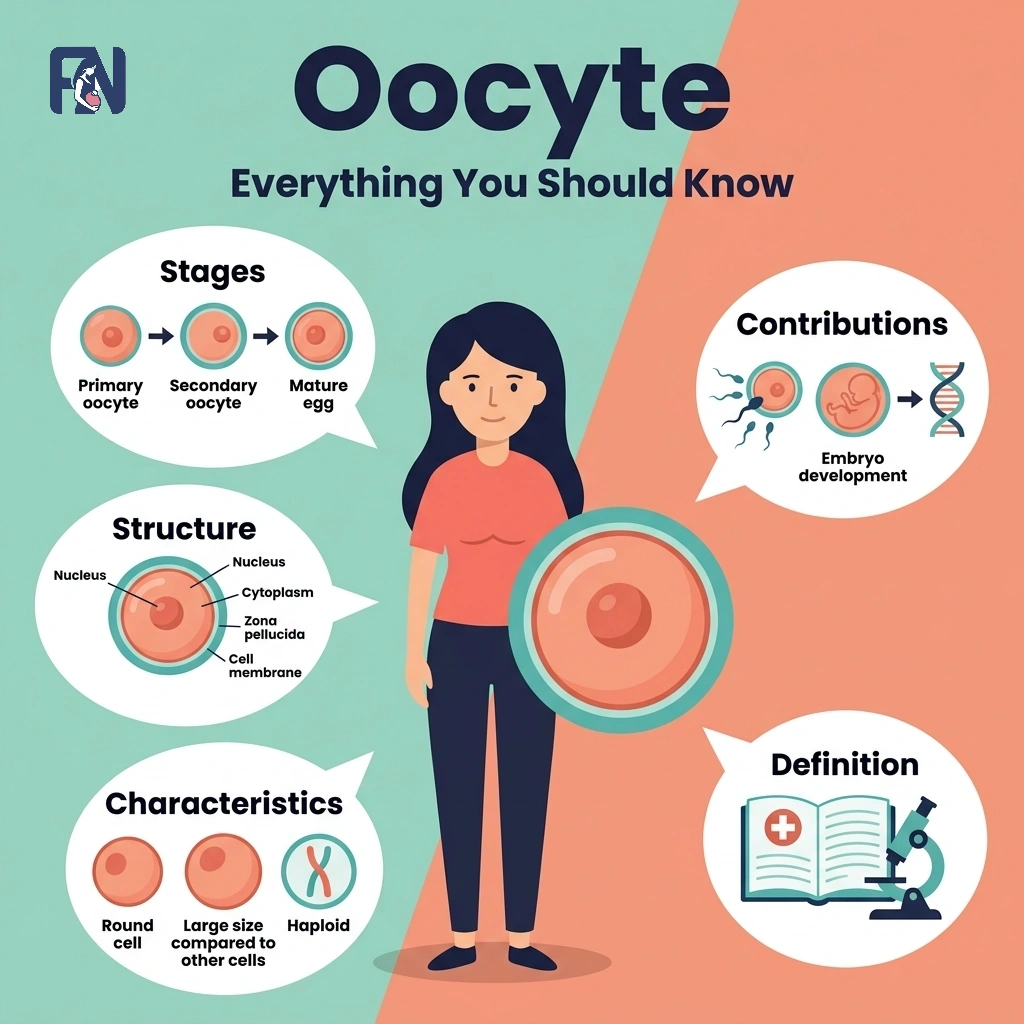
An oocyte refers to an immature female egg cell that plays a central role in reproduction and fertility. Its development spans 4 progressive stages, from primordial germ cells through primary oocytes, secondary oocytes, and finally the mature ovum, each governed by precise hormonal signaling and cellular mechanisms. Structurally, the oocyte includes the nucleus, cytoplasm, zona pellucida, corona radiata, and mitochondria, all of which contribute directly to fertilization competence.
Its characteristics span large cell size, non-renewable supply, arrested meiotic development, high mitochondrial density, and the capacity for cryopreservation through freezing techniques. The oocyte plays a direct role in fertilization by facilitating sperm penetration, completing meiosis II upon sperm entry, and forming the zygote, making it the central biological focus of IVF protocols worldwide. Key factors affecting oocyte health include maternal age, hormonal environment, lifestyle influences, and medical conditions such as endometriosis, each of which is examined in this article.
In India, where infertility affects approximately 10 to 15 percent of couples of reproductive age, understanding oocyte health is a critical component of any fertility assessment. Whether consulting for natural conception challenges or preparing for an IVF cycle, oocyte quality is frequently at the center of the diagnostic and treatment conversation. India now hosts internationally accredited fertility clinics across all major cities, including Mumbai, Delhi, Bangalore, Chennai, and Hyderabad, offering advanced reproductive technologies at a fraction of the cost compared to Western countries. Finding a high-quality IVF center begins with seeking NABH-accredited clinics, reviewing success rates by age group, and consulting a reproductive endocrinologist who specializes in oocyte evaluation and ovarian reserve assessment.
What Is an Oocyte?
An oocyte, also known as an ovocyte, is an immature female germ cell produced within the ovary that undergoes a process called oogenesis to develop into a mature egg capable of fertilization. The oocyte is one of the most biologically complex cells in the human body, designed to carry half the genetic material of future offspring while supplying the cytoplasmic machinery needed to support early embryonic development.
Biologically, the oocyte begins as a primordial germ cell during embryonic development and progresses through distinct stages under hormonal regulation. Its relationship to the ovum is one of developmental progression; the oocyte is the precursor, and the mature egg or ovum represents the final, fertilization-ready form of that cell. The term was first used in scientific literature in the 19th century, with German biologist Oscar Hertwig providing a foundational description of oocyte behavior during fertilization in 1875, marking a turning point in the scientific understanding of the immature female germ cell.
According to the American Society for Reproductive Medicine (ASRM) and data published across National Institutes of Health (NIH) databases, elective oocyte cryopreservation via vitrification results in high survival rates (90%–97%).
Is There a Difference Between an Oocyte and an Egg?
Yes, there is a difference between an oocyte and an egg, as the two terms refer to the same cell at fundamentally different stages of biological development. An oocyte is an immature female reproductive cell that has not yet completed the meiotic divisions required for fertilization, while an egg or ovum is the mature cell capable of being fertilized by a sperm. In the female body, the oocyte develops into a mature egg through the process of maturation and meiosis, culminating in a haploid cell that carries half the chromosomal complement necessary for reproduction.
The distinction matters significantly in clinical settings. During IVF, the oocytes retrieved from follicles are typically at the metaphase II stage, technically classified as secondary oocytes, not fully mature ova, with final meiotic completion triggered only at the moment of fertilization. Knowing this distinction allows fertility specialists to apply the right laboratory conditions, timing protocols, and quality assessment criteria to each developmental stage of the oocyte.
Where Are Oocytes Found?
Oocytes are found in the ovary, specifically within fluid-filled structures called ovarian follicles, which provide the hormonal, nutritional, and structural environment that the oocyte requires to develop and mature through the successive oocyte development stages. In the female body, the ovarian follicle is the primary site for oocyte development because it houses the surrounding granulosa cells and theca cells that produce the estrogen and progesterone necessary for oocyte growth. The proper location of oocytes within follicular microenvironments is essential to the development of a competent, fertilizable egg.
The ovarian reserve, defined as the total pool of remaining ovarian follicles and their enclosed oocytes, diminishes progressively with age. When this reserve becomes significantly depleted, a woman may experience difficulty conceiving, irregular ovulation, or enter into premature ovarian insufficiency or menopause. Monitoring the ovarian reserve through AMH blood tests and antral follicle counts is a fundamental step in fertility evaluation, particularly for women over 30 or those with a history of ovarian surgery or autoimmune conditions.
What Are the Stages of Oocyte Development?
The stages of oocyte development include primordial germ cell formation, primary oocyte arrest in meiosis I, secondary oocyte release at ovulation, and mature ovum formation following fertilization.
The stages of oocyte development are listed below.
- Stage 1 – Primordial Germ Cells: Primordial germ cells refer to the earliest oocyte precursors, specified from epiblast cells and migrating to the developing ovary during weeks 6–8 of gestation. They arise through BMP signaling from the proximal epiblast, migrate via the hindgut and dorsal mesentery under SDF-1 and c-KIT guidance, and colonize the genital ridge, where they undergo mitotic proliferation as oogonia between gestational weeks 6 and 8.
- Stage 2 – Primary Oocyte (Arrest in Meiosis I): The primary oocyte refers to a cell that has entered meiosis I and arrested in prophase I within ovarian follicles, staying dormant from fetal life until puberty. The arrest is maintained by high cAMP and cGMP levels from surrounding granulosa cells; at puberty, the preovulatory LH surge lifts this block, driving the oocyte to complete meiosis I and extrude the first polar body roughly 36 hours before ovulation.
- Stage 3 – Secondary Oocyte (Ovulation Stage): The secondary oocyte is the haploid cell formed when the primary oocyte completes meiosis I, expels the first polar body, and arrests in metaphase II, ready for ovulation. Triggered by the mid-cycle LH surge, it undergoes asymmetric division to preserve cytoplasm, then arrests in metaphase II via cytostatic factor (CSF) until fertilization; ovulation releases it into the fallopian tube about 36–40 hours after the LH peak.
- Stage 4 – Mature Ovum (Post-fertilization Completion): The mature ovum is the fully developed female gamete produced when the secondary oocyte completes meiosis II upon fertilization, expelling the second polar body and forming the haploid maternal pronucleus. Sperm-induced calcium oscillations inactivate cytostatic factor, relieving the metaphase II arrest; meiosis II completes, the second polar body is expelled, and the maternal chromosomes decondense into the female pronucleus, which fuses with the sperm pronucleus to form the zygote within hours of fertilization.
The oocyte development timeline from germ cell migration to zygote formation spans from gestational week 6 through the woman’s reproductive years, with each ovulatory cycle representing one opportunity for a primary oocyte to complete this remarkable structural process.
What Is the Structure of an Oocyte?
The structure of an oocyte includes the nucleus, cytoplasm (ooplasm), zona pellucida, corona radiata, and mitochondria, each performing a specialized biological function that distinguishes the oocyte from a mature ovum and equips it for the demands of fertilization and early embryonic development.
The structural components of an oocyte are listed below.
- Nucleus and Chromosomes: The nucleus refers to the oocyte’s genetic command center, containing 46 chromosomes in the primary stage and 23 chromosomes after meiosis, housing the maternal DNA essential for embryonic inheritance and development.
- Cytoplasm (Ooplasm): The cytoplasm, or ooplasm, refers to the specialized intracellular matrix of the oocyte that houses organelles, maternal mRNA transcripts, and signaling molecules critical for early embryonic development and energy metabolism.
- Zona Pellucida: The zona pellucida refers to a glycoprotein coat encircling the oocyte that mediates sperm binding, supports species-specific fertilization, and triggers the polyspermy block following fertilization, distinguishing the oocyte’s structural role from that of the mature ovum.
- Corona Radiata: The corona radiata refers to the outermost layer of granulosa cells surrounding the zona pellucida, providing nutritional and hormonal support to the oocyte while assisting sperm penetration during fertilization.
- Mitochondria and Energy Role: Mitochondria within the oocyte serve as the primary organelles responsible for energy metabolism, producing the ATP required for meiotic spindle formation, cytoplasmic maturation, and the maintenance of early embryonic development after fertilization.
The integrity of these structural components is a key indicator of oocyte quality in clinical practice.
What Is the Difference Between Oocyte and Ovum?
The difference between an oocyte and ovum is developmental maturity, with an oocyte being an immature germ cell undergoing meiosis and the ovum a mature gamete that has completed meiotic divisions. In terms of oocyte vs ovum characteristics, the oocyte exists across multiple intermediate stages, from prophase I arrest through metaphase II, and is diploid during primary oocyte development, becoming haploid only after completing the first meiotic division. The ovum, by contrast, is a haploid cell structurally primed for sperm fusion and the earliest events of embryonic development.
Understanding this distinction is directly relevant to fertility tracking and IVF procedures. During oocyte retrieval in an IVF cycle, the cells collected from follicles are metaphase II oocytes, not yet full ova, and full meiotic maturation is completed only after successful fertilization. Clinicians use this distinction to design insemination timing strategies, evaluate oocyte maturity rates after retrieval, and select the most biologically advanced cells for fertilization and embryo culture.
What Are the Characteristics of Oocytes?
The characteristics of oocytes include their large cell size, arrested meiotic development, non-renewable supply, high mitochondrial content, zona pellucida enclosure, and capacity for cryopreservation through freezing, distinguishing them as the most biologically complex and specialized cells in the female reproductive system.
The characteristics of oocytes are listed below.
- Large Cell Size: Oocytes are among the largest cells in the human body, measuring approximately 100 to 120 micrometers in diameter, enabling sufficient storage of cytoplasmic reserves for early embryo development before implantation.
- Arrested Meiotic Development: Oocytes remain arrested in prophase I of meiosis I from fetal life until puberty, resuming division only in response to a pre-ovulatory LH surge, a feature unique to female germ cell biology.
- Non-Renewable Supply: Unlike sperm, oocytes are produced only during fetal development and cannot be regenerated after birth, making ovarian reserve preservation and monitoring essential to long-term reproductive health.
- High Mitochondrial Density: Oocytes contain more mitochondria than virtually any other cell type in the human body, reflecting their extraordinary energy demands during meiotic maturation, fertilization, and early embryonic cleavage.
- Zona Pellucida Enclosure: Oocytes are encased in the zona pellucida, a glycoprotein shell that governs sperm binding specificity, protects genetic material, and coordinates the polyspermy block after fertilization.
- Capacity for Cryopreservation: Oocytes can be preserved long-term through the freezing technique known as vitrification, with post-thaw survival rates exceeding 80% in experienced centers, enabling women to store reproductive potential for future use.
These characteristics define the oocyte’s unique biological identity and are directly relevant to how fertility specialists evaluate and manage egg quality during clinical fertility assessments and treatment planning cycles.
Is It Possible to Freeze Oocytes?
Yes, it is possible to freeze oocytes (eggs), and oocyte cryopreservation is now considered a well-established, safe, and clinically effective procedure in reproductive medicine. The procedure is clinically known as oocyte cryopreservation and involves collecting unfertilized eggs from the ovaries following a course of controlled ovarian stimulation, after which they are vitrified, rapidly cooled at ultra-high speeds to prevent ice crystal formation and preserve cellular integrity for long-term storage.Oocyte health at the time of retrieval is the most critical determinant of post-thaw survival and subsequent fertilization success
What Factors Affect Oocyte Health?
The factors that affect oocyte health include maternal age, hormonal environment, lifestyle influences, medical conditions such as endometriosis, oxidative stress, and genetic background.
The factors affecting oocyte health are listed below.
- Maternal Age: Maternal age is the single most significant factor affecting oocyte quality, as advancing age accelerates the accumulation of chromosomal aneuploidy errors during meiosis, sharply increasing the risk of failed fertilization and miscarriage after 35.
- Hormonal Environment: The hormonal environment, comprising FSH, LH, estrogen, and AMH levels, directly governs follicular development and oocyte maturation, and imbalances caused by PCOS, hypothyroidism, or hyperprolactinemia can impair egg quality significantly.
- Lifestyle Influences: Lifestyle factors including smoking, alcohol consumption, poor diet, obesity, and high chronic stress reduce mitochondrial efficiency, increase oocyte DNA damage, and impair the hormonal signaling needed to improve oocyte quality before retrieval.
- Medical Conditions: Conditions such as endometriosis, polycystic ovary syndrome, thyroid disorders, and autoimmune disease can disrupt the follicular microenvironment and reduce both the quantity and quality of retrievable oocytes during stimulation.
- Oxidative Stress: Elevated reactive oxygen species in follicular fluid create an oxidative environment that damages oocyte DNA, destabilizes meiotic spindle assembly, and diminishes the oocyte’s capacity to support successful fertilization and embryo development.
- Genetic Factors: Heritable mutations in genes regulating meiotic fidelity, mitochondrial biogenesis, or ovarian reserve maintenance can predispose women to early oocyte depletion, poor morphology, and reduced fertilization rates independent of lifestyle or age.
What Is the Role of the Oocyte in Fertilization?
The role of the oocyte in fertilization includes facilitating sperm penetration through the zona pellucida, resuming and completing meiosis II upon sperm entry, triggering the cortical reaction to prevent polyspermy, and enabling the formation of a viable zygote. These functions define the oocyte fertilization role and establish it as an active, rather than passive, participant in the process of conception.
When a sperm cell penetrates the zona pellucida and fuses with the oocyte’s plasma membrane, it delivers the biochemical signal that triggers meiosis II completion. This division produces the second polar body and yields the female pronucleus. The male pronucleus, delivered by the fertilizing sperm, fuses with the female pronucleus to complete zygote formation, the first cell of a new organism. The entire sequence from sperm penetration to zygote formation occurs over a span of approximately 12 to 20 hours following sperm-egg fusion.
The cortical reaction is a critical oocyte-driven response in which cortical granules beneath the plasma membrane release enzymes into the perivitelline space immediately after sperm fusion. These enzymes chemically modify the zona pellucida, rendering it impenetrable to additional sperm, a mechanism known as the polyspermy block. This reaction is essential to ensure the embryo develops from a diploid, single-sperm fertilization, as polyspermy produces chromosomally abnormal, non-viable embryos. In clinical IVF settings, the integrity of this oocyte mechanism is assessed by confirming the presence of two pronuclei 16 to 18 hours after insemination.
What Are the Common Problems Affecting Oocytes?
The common problems affecting oocytes include several developmental and chromosomal conditions that can impair fertilization, embryo viability, and overall reproductive success. The common problems affecting oocytes are listed below.
- Poor Oocyte Quality: Poor oocyte quality refers to morphological or cytoplasmic abnormalities, such as granular cytoplasm, vacuolation, or zona pellucida irregularities, that reduce fertilization and embryo development potential.
- Chromosomal Abnormalities: Chromosomal abnormalities refer to aneuploidies arising from meiotic division errors in the oocyte, leading to failed fertilization, early pregnancy loss, or chromosomally affected offspring.
- Diminished Ovarian Reserve: Diminished ovarian reserve refers to a reduced pool of remaining follicles and oocytes, resulting in fewer viable eggs available for natural ovulation or IVF retrieval and a lower likelihood of conception.
- Anovulation: Anovulation refers to cycles in which no mature oocyte is released from the ovary, preventing fertilization entirely and requiring medical intervention as a first step in treating the fertility issues it causes.
- Oocyte Fragmentation: Oocyte fragmentation refers to cytoplasmic or zona pellucida irregularities that compromise oocyte structural integrity, reducing success rates in both natural fertilization and assisted reproductive technology cycles.
Accurate diagnosis of these problems requires a comprehensive fertility workup including AMH testing, antral follicle count, hormonal profiling, and oocyte morphology assessment. Early identification allows fertility specialists to select the most appropriate treatment pathway for each patient’s individual clinical profile.
What Are the Treatments for Poor Oocyte Quality?
The treatment options for poor oocyte quality involve a combination of evidence-based lifestyle changes, medical protocols, and advanced assisted reproductive technologies designed to optimize the follicular environment and improve the developmental competence of retrieved eggs.
The treatments for poor oocyte quality are listed below.
- Lifestyle Modifications: Lifestyle modifications refer to evidence-based changes in diet, exercise, sleep hygiene, and stress management that reduce oxidative stress and improve mitochondrial function, supporting a more favorable environment for oocyte development before retrieval.
- Custom IVF Protocols: Custom IVF protocols refer to individualized ovarian stimulation regimens using FSH, LH, and GnRH analogues tailored to the patient’s hormonal profile, aiming to retrieve the maximum number of competent mature oocytes per cycle.
- In Vitro Maturation (IVM): In Vitro Maturation refers to an evidence-based fertility technique in which immature oocytes are collected from unstimulated or minimally stimulated ovaries and cultured in the laboratory to the metaphase II stage before fertilization.
- Preimplantation Genetic Testing (PGT): Preimplantation Genetic Testing refers to a biopsy-based chromosomal screening procedure performed on embryos derived from retrieved oocytes to identify euploid embryos before transfer, improving implantation and live birth rates.
- Platelet-Rich Plasma (PRP) Therapy: Platelet-Rich Plasma therapy refers to an evidence-based ovarian rejuvenation treatment in which autologous PRP is injected into ovarian tissue to stimulate follicular activation and potentially enhance oocyte quality in women with diminished ovarian reserve.
- Artificial Oocyte Activation (AOA): Artificial Oocyte Activation refers to a laboratory technique that uses calcium ionophore or mechanical means to mimic sperm-triggered calcium oscillations, applied in ICSI failure cases to restore normal oocyte activation and improve fertilization rates.
- Supplements: Supplements refer to evidence-based micronutrients and antioxidants,, including CoQ10, melatonin, DHEA, vitamin D, and omega-3 fatty acids, used to reduce oxidative damage and support mitochondrial energy metabolism in oocytes during the pre-treatment period.
Many leading Indian fertility centers now offer the full spectrum of these interventions within integrated reproductive medicine programs.
What Are the Best Places in India for Oocyte Abnormality Treatment?
The best places in India for oocyte and fertility treatment include major metropolitan areas such as Mumbai, Delhi NCR, Bangalore, Chennai, Hyderabad, Kolkata, and Ahmedabad, all of which host internationally accredited fertility clinics and IVF centers equipped with advanced embryology laboratories and experienced reproductive medicine teams.
The best places in India for fertility treatment are listed below.
- Mumbai, Maharashtra: Mumbai houses some of India’s most established fertility centers, including institutions led by specialists offering advanced oocyte retrieval, IVF, and ICSI for complex oocyte abnormality cases.
- Delhi: Delhi is home to a dense network of reproductive medicine hospitals offering custom IVF protocols, PRP therapy, PGT, and AI-assisted embryo selection, supported by experienced teams specializing in complex oocyte-related fertility conditions.
- Bangalore, Karnataka: Bangalore hosts internationally accredited fertility centers combining modern IVF technology with comprehensive oocyte quality optimization programs and advanced embryology services, catering to both domestic patients and medical tourists.
- Chennai, Tamil Nadu: Chennai offers high-success-rate IVF programs within well-equipped tertiary hospitals and dedicated fertility clinics providing IVM, PGT, and advanced embryology services for oocyte abnormality evaluation and treatment.
- Hyderabad, Telangana: Hyderabad has rapidly developed its fertility infrastructure, with specialized centers providing AI-assisted oocyte and embryo selection, oocyte cryopreservation, and individualized stimulation protocols at competitive Indian prices.
- Kolkata, West Bengal: Kolkata provides accessible fertility treatment in eastern India, with IVF centers offering a range of oocyte-related interventions, including cryopreservation supported by experienced reproductive medicine specialists.
- Ahmedabad, Gujarat: Ahmedabad is emerging as a significant fertility treatment hub in western India, with clinics offering oocyte cryopreservation, IVF, ICSI, and integrative reproductive care combining modern medicine with complementary wellness approaches.
India’s fertility treatment landscape has expanded substantially over the last decade, with NABH-accredited clinics in these cities delivering outcomes comparable to international standards while remaining significantly more affordable than equivalent care in the US, UK, or Australia.
How Can Modern Indian Treatments Help with Oocyte-Related Infertility?
Modern Indian treatments can help address oocyte-related infertility by improving egg quality, supporting ovarian function, and increasing the chances of successful conception through a broad array of advanced reproductive technologies and individualized, patient-centered care models.
In terms of assisted reproductive technologies, Indian fertility centers now offer the full spectrum of evidence-based infertility treatments, including IVF, ICSI, and IVM, all of which are designed to work around limitations in oocyte quantity or quality and improve fertilization and embryo development outcomes.
IVF protocols are increasingly customized to individual hormonal profiles and ovarian reserve assessments, allowing clinicians to optimize stimulation outcomes and retrieve the most viable oocytes possible. Time-lapse embryo monitoring and AI-assisted egg and embryo selection are also now available at leading Indian centers, enabling more precise identification of the most developmentally competent oocytes and resulting embryos for transfer.
Emerging and supportive therapies available in India include PRP ovarian rejuvenation for women with diminished ovarian reserve, artificial oocyte activation for ICSI failure cases, and PGT to identify chromosomally normal embryos. These innovations allow Indian fertility specialists to manage complex oocyte abnormality cases with high precision and meaningful clinical success rates.
A distinctive aspect of the Indian fertility care landscape is the availability of integrative treatment models that combine modern reproductive medicine with supportive Ayurvedic therapies.
How Much Does Oocyte Abnormality Treatment Cost in India?
Oocyte abnormality treatment in India typically costs between ₹1,00,000 and ₹4,50,000 per cycle, depending on the clinic, protocol, and medication requirements. This makes Indian fertility care substantially more affordable than equivalent treatments in Western countries while maintaining internationally comparable success rates in accredited centers.
The standard cost of treatment generally includes the core IVF or ICSI procedure, ovarian stimulation monitoring through ultrasound and blood tests, oocyte retrieval and laboratory embryology services, and basic embryo culture and transfer. In cases requiring additional procedures, such as oocyte cryopreservation, PGT, IVM, or PRP ovarian therapy, the per-cycle cost may rise to ₹3,00,000 or above, depending on the specific techniques used and the patient’s individual clinical requirements.
Several factors affect the final cost, including the city and specific clinic chosen, the complexity of the stimulation protocol designed around individual hormonal profiles and ovarian reserve measurements, the type and dosage of fertility medications prescribed. Premium accredited fertility centers in Mumbai and Delhi typically charge higher fees than well-equipped clinics in Hyderabad or Ahmedabad, though accredited centers across these cities generally deliver comparable success rates for most oocyte-related conditions.
Oocyte abnormality cases involving the different oocyte stages, including immature oocytes requiring IVM or cases of severely diminished ovarian reserve, may require multiple stimulation cycles or advanced add-on procedures, increasing the overall treatment investment. Patients seeking to understand the roles in fertilization of oocyte quality, how the oocyte development timeline and proper ovocyte structure influence treatment success, or which specific treatments for poor oocyte quality best match their clinical profile, are strongly encouraged to consult a board-certified reproductive endocrinologist for a personalized evaluation.
To explore oocyte health assessment and fertility treatment options tailored to your specific needs, visit Fertility Network for a comprehensive directory of accredited IVF centers, expert consultations, and patient support resources across major Indian cities.

Fathima Zahra is a reproductive health content specialist at FertilityNetwork.in — India’s largest independent fertility clinic directory and patient education platform.
Fathima’s work covers the full spectrum of reproductive medicine, from foundational biology to advanced fertility treatment. Her published articles span reproductive anatomy (ovaries, uterus, fallopian tubes, testes), core reproductive processes (ovulation, fertilization, implantation, the menstrual cycle), reproductive hormones (estrogen, progesterone, testosterone, FSH, LH, AMH, and prolactin), and the clinical conditions that cause infertility in both men and women.
On the male side, her research covers conditions including azoospermia, oligospermia, asthenozoospermia, teratozoospermia, varicocele, erectile dysfunction, Klinefelter syndrome, and Y chromosome microdeletion. On the female side, she writes about PCOS, endometriosis, uterine fibroids, anovulation, diminished ovarian reserve, premature ovarian insufficiency, tubal blockage, pelvic inflammatory disease, and Turner syndrome.
Her treatment coverage includes intrauterine insemination (IUI), in vitro fertilization (IVF), intracytoplasmic sperm injection (ICSI), egg retrieval, embryo transfer, egg freezing, sperm freezing, and oncofertility — providing patients with detailed, process-level understanding of each procedure, its clinical indications, success rates, and costs.
Every article Fathima publishes follows a structured, evidence-based research methodology:
1. Source verification against ICMR (Indian Council of Medical Research) national fertility and ART guidelines
2. Cross-referencing with ESHRE (European Society of Human Reproduction and Embryology) clinical protocols
3. Alignment with ASRM (American Society for Reproductive Medicine) evidence-based practice guidelines
4. Citation of PubMed-indexed, peer-reviewed studies published within the last 5 years
5. Verification of all diagnostic thresholds, hormonal reference ranges, and clinical statistics against current international standards
All content authored by Fathima undergoes editorial review by the FertilityNetwork Medical Research Board before publication, ensuring clinical accuracy and factual integrity across every article published on the platform.
Fathima’s content is editorially independent. FertilityNetwork.in does not publish sponsored medical opinions, and no article is written to promote specific clinics, doctors, or fertility treatments.
Leave a Reply