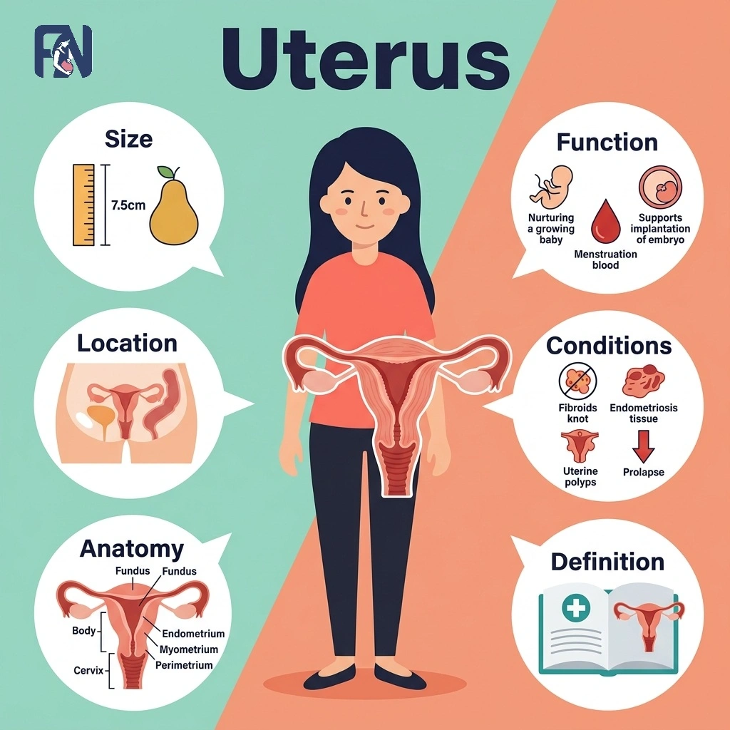
The uterus refers to the muscular organ in the female pelvis where a fetus develops during pregnancy. In a non-pregnant adult of reproductive age, it measures about 7 to 8 centimeters long and sits in the pelvis between the bladder and rectum, connected to the fallopian tubes, cervix, and vagina. Its anatomy includes distinct regions such as the fundus, body, isthmus, and cervix, along with three tissue layers that support menstruation, implantation, and pregnancy.
Understanding how the uterus functions and how hormones like estrogen, progesterone, and oxytocin regulate it helps explain everything from a typical menstrual cycle to labor and delivery. Some common uterine conditions are fibroids, adenomyosis, endometriosis, polyps, uterine prolapse, endometrial hyperplasia, and uterine cancer, along with the symptoms that can signal a problem.
Monitoring uterine health through regular pelvic exams and timely assessment of symptoms such as heavy bleeding or pelvic pain supports early diagnosis, and treatment options range from medication and hormone therapy to minimally invasive procedures, myomectomy, or hysterectomy depending on the condition.
What Is the Uterus?
The uterus is a hollow, muscular reproductive organ located in the female pelvis that houses and nourishes a developing embryo and fetus during gestation. The uterus is commonly called the womb because it serves as the site where a fertilized egg implants and grows throughout pregnancy.
Pear-shaped and firm yet flexible in consistency, the uterus is a hormone-responsive organ that changes in size and structure in response to estrogen and progesterone. This responsiveness underlies its central reproductive significance, from monthly menstrual cycles to supporting a full-term pregnancy.
What Is the Normal Size of the Uterus?
The normal size of the uterus is about 7–8 cm (2.8–3 inches) long, 4–5 cm (1.6–2 inches) wide, and 2–3 cm (0.8–1.2 inches) thick in a non-pregnant adult of reproductive age. It is typically pear-shaped and weighs approximately 60 grams, with its exact locational position and size varying slightly from person to person. Age, hormonal changes, the phase of the menstrual cycle, and childbirth history can all influence uterine size, with the uterus often appearing slightly larger in women who have given birth.
Where Is the Uterus Located?
The uterus is located in the center of the female pelvis, positioned between the bladder in front and the rectum behind. This anatomical position allows the uterus to connect directly with the fallopian tubes at its upper corners and with the cervix and vagina below.
The uterus is typically tilted forward, a position known as anteversion, though a backward tilt, or retroversion, is also common and not considered abnormal. Uterine position can vary among individuals without indicating any underlying problem.
What Is the Anatomy of the Uterus?
The anatomy of the uterus includes the fundus, body (or corpus), isthmus, and cervix. Each region and layer plays a specific role in supporting menstruation, implantation, and pregnancy.
The key anatomical structures of the uterus are listed below.
- Fundus: The fundus is the rounded, superior portion of the uterus, located above where the fallopian tubes attach on either side. It forms the top of the uterine body and is the widest part of the organ. During pregnancy, doctors measure fundal height from this point to track fetal growth and estimate gestational age.
- Body (Corpus): The body, or corpus, of the uterus refers to the main, central portion of the organ between the fundus and the isthmus. It is the largest part of the uterus and contains the uterine cavity, the site where implantation occurs. The corpus expands dramatically during pregnancy to accommodate the growing fetus.
- Isthmus: The isthmus is the narrow region of the uterus located between the body and the cervix. During pregnancy, this area stretches and thins to form the lower uterine segment. Because it thins predictably, the isthmus is also the standard incision site during a cesarean section.
- Cervix: The cervix refers to the lower, narrow portion of the uterus that connects the uterine cavity to the vagina through the cervical canal. Its inner layer produces cervical mucus, which changes in texture throughout the menstrual cycle to support or block sperm passage. The cervix also plays a critical function during childbirth, dilating fully to allow delivery.
What Are the Layers of the Uterine Wall?
The 3 layers of the uterine wall are listed below.
- Endometrium: The endometrium is the innermost layer of the uterine wall, made up of a functional layer that sheds monthly and a basal layer that regenerates it. Hormonal changes in estrogen and progesterone drive the functional layer’s growth and shedding during the menstrual cycle. Endometrial glands within this layer support implantation when a fertilized egg arrives.
- Myometrium: The myometrium refers to the thick, middle layer of smooth muscle that makes up most of the uterine wall. It enables the uterus to enlarge dramatically during pregnancy and to generate the powerful contractions needed for labor, triggered by oxytocin. After childbirth, the myometrium undergoes postpartum involution, gradually shrinking the uterus back toward its pre-pregnancy size.
- Perimetrium: The perimetrium refers to the outer serosal layer that covers the uterus, formed by the visceral peritoneum. It connects the uterus to surrounding pelvic structures and helps anchor its position within the pelvis. This outer layer also contains nerve fibers that contribute to pelvic sensation.
What Are the Main Components of Uterine Blood Supply, Lymphatics, and Innervation?
The main components of uterine blood supply, lymphatics, and innervation are listed below.
- Arterial Supply: The arterial supply of the uterus refers to the network of blood vessels that deliver oxygenated blood to support its function. The uterine artery, a branch of the internal iliac artery, provides the main blood supply, while the ovarian artery contributes additional flow near the fundus. Together, these vessels ensure the uterus receives adequate blood throughout the menstrual cycle and pregnancy.
- Venous Drainage: Venous drainage of the uterus refers to the system of veins that carry deoxygenated blood away from the organ. The uterine vein serves as the primary vessel for this drainage, running alongside the uterine artery before emptying into the internal iliac vein. Efficient venous drainage helps prevent congestion and supports healthy uterine tissue.
- Lymphatic Drainage: Lymphatic drainage of the uterus is the network of vessels that carry lymph fluid away from uterine tissue toward the pelvic lymph nodes. This drainage supports immune function and helps clear waste products from the uterine wall. Because uterine cancer can spread through this pathway, pelvic lymph nodes are closely examined during cancer staging.
- Nerve Supply: The nerve supply of the uterus refers to the network of autonomic nervous system fibers that regulate uterine sensation and muscle activity. These nerves largely originate from the uterovaginal plexus, which carries both sympathetic and parasympathetic input. This nerve supply influences functions such as pain perception and the coordination of contractions during labor.
What Are the Functions of the Uterus?
The functions of the uterus include menstruation, implantation, supporting pregnancy, and enabling labor. These functions work together to make conception, pregnancy, and childbirth possible.
The main functions of the uterus are listed below.
- Menstruation: Menstruation is the monthly shedding of the uterine lining, or endometrium, when pregnancy does not occur. The uterus initiates menstruation by shedding its inner lining, the endometrium, when fertilization does not occur. This cyclical process reflects the uterus’s role in hormonal regulation and preparation for potential pregnancy.
- Implantation: Implantation is the process by which a fertilized egg, now a blastocyst, attaches to the uterine lining about six to ten days after fertilization. Once attached, the blastocyst begins burrowing into the endometrium and initiates the early stages of placental development. Successful implantation marks the true biological start of pregnancy.
- Pregnancy: The uterus supports pregnancy by expanding dramatically in size to accommodate the growing fetus, from roughly the size of a fist to that of a watermelon. Its muscular walls protect the fetus from outside pressure and impact throughout gestation. Through the placenta, the uterus also facilitates nutrient and oxygen exchange between mother and baby.
- Labor: The uterus’s role in labor involves powerful myometrial contractions triggered by rising oxytocin levels as childbirth approaches. These contractions gradually cause cervical dilation, widening the cervix to allow the baby to pass through the birth canal. This hormonal and mechanical process culminates in delivery of the baby and, shortly after, the placenta.
What Are the Hormonal Regulation of the Uterus?
The hormones that regulate the uterus include estrogen, progesterone, LH, FSH, oxytocin, and prostaglandins.
The hormones that regulate the uterus are listed below.
- Estrogen: Estrogen is the hormone that binds to hormone receptors in the uterine lining to drive its growth. It plays a central role in endometrial proliferation during the first half of the menstrual cycle.
- Progesterone: Progesterone is the hormone released after ovulation that maintains the secretory phase of the endometrium. This phase thickens and stabilizes the uterine lining to support implantation if fertilization occurs.
- LH: LH, or luteinizing hormone, is the pituitary hormone that surges midcycle to trigger ovulation. It also supports corpus luteum function afterward, which sustains progesterone production for the uterine lining.
- FSH: FSH, or follicle-stimulating hormone, refers to the pituitary hormone that stimulates follicle development in the ovaries. By promoting follicle growth, FSH indirectly supports the estrogen levels that shape uterine hormone activity.
- Oxytocin: Oxytocin is the hormone released by the pituitary gland that triggers myometrial contractions during labor. In the uterus, oxytocin released by the pituitary gland stimulates the muscular layer (myometrium) to contract during labor. Its levels rise sharply as delivery approaches, coordinating the rhythmic contractions needed for childbirth.
- Prostaglandins: Prostaglandins are hormone-like compounds that trigger uterine contractions involved in menstrual shedding and, later in life, cervical ripening before labor. These compounds play a role in uterine function throughout a woman’s lifetime, from her first period through childbirth.
How Does the Uterus Change Throughout Life?
The 5 life stages of uterine change are listed below.
- Childhood: During childhood, the uterus remains small and structurally immature, reflecting typical prepubertal uterus anatomy. Its reproductive function stays inactive until hormonal changes begin later in adolescence.
- Puberty: During puberty, the uterus undergoes hormonal maturation as rising estrogen levels stimulate its growth. This stage culminates in menarche, the first menstrual period, marking the start of reproductive maturity.
- Reproductive Years: During the reproductive years, the uterus undergoes monthly cycles of lining growth and shedding tied to ovulation and the menstrual cycle. These cycles prepare the uterus each month to support fertility and potential pregnancy.
- Pregnancy: During pregnancy, the uterus enlarges dramatically, with increased blood flow supporting placenta formation and ongoing fetal development. It stretches from the size of a fist to fill much of the abdominal cavity by full term.
- Menopause: During menopause, the uterus responds to hormonal decline and estrogen deficiency with endometrial thinning and gradual uterine atrophy. Maintaining uterine health during this stage still matters, since postmenopausal bleeding can signal an underlying problem.
How to Maintain Uterine Health?
The habits that support uterine health are listed below.
- Schedule Regular Gynecological Exams: Regular gynecological exams, including a pelvic examination, allow doctors to detect uterine conditions early, often before symptoms appear.
- Get Cervical Cancer Screening: Cervical cancer screening through a Pap smear checks for abnormal cervical cells and is generally recommended every three years for most women.
- Maintain a Healthy Weight: Maintaining a healthy weight supports balanced hormone levels, since excess body fat can disrupt estrogen levels and increase uterine health risks.
- Stay Physically Active: Regular physical activity supports hormonal balance and healthy blood flow, both of which benefit reproductive health over time.
- Practice STI Prevention: STI prevention, including consistent condom use, helps protect against infections that can lead to pelvic inflammatory disease and uterine damage.
- Quit Smoking: Smoking cessation reduces the risk of several uterine conditions, including cervical cancer and fertility complications linked to reduced blood flow.
- Attend Prenatal Care Visits: Prenatal care during pregnancy allows doctors to monitor uterine and fetal health, catching complications early for a safer pregnancy.
- Get the HPV Vaccination: HPV vaccination protects against the human papillomavirus strains most commonly linked to cervical cancer and is recommended before or during early adulthood as part of a healthy lifestyle.
What Are the Common Uterine Conditions?
The common uterine conditions include fibroids, adenomyosis, endometriosis, polyps, uterine prolapse, endometrial hyperplasia, and uterine cancer, each affecting the uterus differently and requiring distinct treatment approaches.
The common uterine conditions are listed below.
- Fibroids: Fibroids refer to noncancerous growths that develop within or on the walls of the uterus, varying widely in size and number. They are commonly linked to estrogen sensitivity, genetics, and family history, with risk increasing with age until menopause. Common symptoms include heavy menstrual bleeding, pelvic pressure, and, in some cases, complications with fertility.
- Adenomyosis: Adenomyosis is a condition in which endometrial tissue grows into the myometrium, the muscular wall of the uterus. Its exact cause is unclear, though risk factors include prior uterine surgery and childbirth. Common symptoms include painful, heavy periods and an enlarged, tender uterus.
- Endometriosis: Endometriosis is a condition in which tissue similar to the uterine lining grows outside the uterus, often on the ovaries or pelvic tissue. Its causes remain unclear, though genetics and retrograde menstruation are considered contributing risk factors. Common symptoms include pelvic pain and infertility, which can range from mild to severe.
- Polyps: Uterine polyps refer to small, usually noncancerous growths that develop from the endometrial lining and extend into the uterine cavity. They are linked to excess estrogen exposure and become more common with age, particularly around menopause. The most common symptom is abnormal bleeding, including spotting between periods.
- Uterine Prolapse: Uterine prolapse refers to a condition in which the uterus slips down into or out of the vaginal canal due to weakened support structures. It is commonly caused by pelvic floor weakness following childbirth, aging, or chronic straining. Symptoms often include a sensation of pelvic heaviness or a visible bulge at the vaginal opening.
- Endometrial Hyperplasia: Endometrial hyperplasia is abnormal thickening of the uterine lining, usually caused by excess estrogen without enough progesterone to balance it. Risk factors include obesity, polycystic ovary syndrome, and irregular ovulation. The most common symptom is abnormal uterine bleeding, which requires evaluation since it can sometimes precede cancer.
- Uterine Cancer: Uterine cancer refers to a malignant growth that typically begins in the endometrial lining of the uterus. Risk factors include obesity, prolonged estrogen exposure, and a family history of the disease. Common symptoms include abnormal vaginal bleeding, particularly after menopause, along with pelvic pain in more advanced cases.
What Symptoms Can Indicate a Uterine Problem?
The common symptoms that may indicate a uterine problem are listed below.
- Heavy Menstrual Bleeding: Heavy menstrual bleeding refers to periods with unusually heavy flow or prolonged duration. It can be a sign of fibroids, polyps, or hormonal imbalance.
- Pelvic Pain: Pelvic pain refers to discomfort in the lower abdomen that can range from mild to severe. It can result from fibroids, adenomyosis, or endometriosis.
- Infertility: Infertility refers to the inability to conceive after a year of regular, unprotected intercourse. Structural or hormonal uterine problems can prevent conception by disrupting implantation.
- Abnormal Bleeding: Abnormal bleeding refers to vaginal bleeding that occurs outside a normal menstrual pattern. It includes bleeding between periods or after intercourse and may signal an underlying condition.
- Postmenopausal Bleeding: Postmenopausal bleeding refers to any vaginal bleeding that occurs after menopause has been confirmed. It always requires prompt medical evaluation to rule out uterine cancer.
- Pelvic Pressure: Pelvic pressure means a heavy or full sensation in the lower abdomen or pelvis. It is commonly associated with fibroids or uterine prolapse.
- Painful Intercourse: Painful intercourse refers to discomfort or sharp pain felt during or after sexual activity. It can result from endometriosis, adenomyosis, or pelvic inflammation.
- Abnormal Discharge: Abnormal discharge means vaginal discharge that differs in color, odor, or consistency from what is typical. It may signal infection, polyps, or, rarely, uterine cancer.
- Recurrent Miscarriage: Recurrent miscarriage refers to the loss of two or more consecutive pregnancies before 20 weeks. Uterine abnormalities, such as a septum or fibroids, can be an underlying cause.
How Are Uterine Conditions Diagnosed?
The common methods used to diagnose uterine conditions are listed below.
- Pelvic Examination: A pelvic examination is a physical exam in which a doctor checks the uterus, cervix, and surrounding structures. It includes a bimanual examination and a speculum examination to assess size, shape, and tenderness.
- Ultrasound: An ultrasound, including transabdominal and transvaginal ultrasound, is an imaging test that uses sound waves to create images of the uterus. It helps visualize the uterus and detect abnormalities such as fibroids, polyps, or thickened lining.
- MRI: A pelvic MRI is an imaging test that uses magnetic fields to create highly detailed pictures of pelvic structures. It provides detailed soft tissue imaging of the uterus, helping distinguish between different types of masses.
- CT Scan: A CT scan, or computed tomography, is an imaging test that combines multiple X-ray images to create cross-sectional pictures of the pelvis. It provides pelvic imaging often used to assess whether uterine cancer has spread.
- Hysteroscopy: Hysteroscopy is a procedure in which a thin, lighted scope is inserted through the cervix into the uterus. It allows direct visualization of the uterine cavity to detect and sometimes treat polyps or fibroids.
- Endometrial Biopsy: An endometrial biopsy is a procedure that involves taking a small tissue sample from the uterine lining. This sample undergoes histopathology to detect abnormal cells and helps guide decisions about appropriate treatments.
What Are the Best Treatments for Uterine Conditions in India?
The best treatments for uterine conditions in India include medications, hormone therapy, minimally invasive procedures, myomectomy, and hysterectomy, available at major hospitals across the country.
The best treatments for uterine conditions in India are listed below.
- Delhi: Delhi offers advanced gynecological centers providing medications, hormone therapy, and minimally invasive procedures such as laparoscopic myomectomy, with treatment cost-range varying by hospital and procedure.
- Mumbai: Mumbai is home to leading hospitals performing myomectomy and hysterectomy alongside fertility-focused uterine care, with a wide cost range depending on complexity.
- Bangalore: Bangalore provides access to gynecologists specializing in minimally invasive procedures and hormone therapy for conditions such as fibroids and endometriosis.
- Chennai: Chennai has established centers offering both medical management and surgical treatments, including myomectomy and hysterectomy, for a range of uterine conditions.
- Hyderabad: Hyderabad offers well-equipped hospitals providing hormone therapy and minimally invasive procedures, with treatment costs varying based on diagnosis and approach.
- Kolkata: Kolkata has growing gynecological infrastructure with specialists treating fibroids, adenomyosis, and other uterine conditions through medication or surgery.
- Pune: Pune provides access to gynecological specialists and diagnostic centers offering treatment options ranging from medication to hysterectomy, depending on condition severity.
How Much Does Uterine Condition Treatment Cost in India?
The cost of uterine condition treatment in India ranges from ₹15,000 to ₹4,00,000 depending on the diagnosis, treatment approach, and hospital chosen. Initial consultations and diagnostic tests generally cost between ₹500 and ₹2,500 per visit. Treatment costs vary further by approach, with medication and hormone therapy at the lower end and minimally invasive procedures, myomectomy, or hysterectomy at the higher end.
Can Uterine Conditions Cause Infertility?
Yes, uterine conditions can cause infertility because they can disrupt implantation, hormone balance, or the shape of the uterine cavity needed for conception. These conditions interfere with the uterus’s ability to support a fertilized egg or maintain a healthy pregnancy. Common underlying conditions associated with infertility include fibroids, adenomyosis, endometriosis, and polyps, all of which can directly affect reproductive health.
How to Find the Right Treatments in India for Infertility Caused by Uterine Conditions?
To find the right infertility treatment in India for uterine conditions, compare experienced fertility specialists, recommended treatments, and treatment costs based on your diagnosis and reproductive needs.
The right treatment depends on the anatomy of the uterus, its functions, hormonal regulation, the size and location of the uterus, and the specific uterine conditions affecting fertility. Important treatments include medications, hysteroscopic surgery, myomectomy, treatment for uterine polyps or adhesions, and fertility treatments such as IUI or IVF when recommended.
Before choosing a clinic, check the accuracy of the diagnosis, specialist expertise, success rates, available facilities, follow-up care, and treatment cost. Fertility Network helps you find affordable fertility treatment options near you in India by connecting you with trusted fertility specialists and clinics.

Fathima Zahra is a reproductive health content specialist at FertilityNetwork.in — India’s largest independent fertility clinic directory and patient education platform.
Fathima’s work covers the full spectrum of reproductive medicine, from foundational biology to advanced fertility treatment. Her published articles span reproductive anatomy (ovaries, uterus, fallopian tubes, testes), core reproductive processes (ovulation, fertilization, implantation, the menstrual cycle), reproductive hormones (estrogen, progesterone, testosterone, FSH, LH, AMH, and prolactin), and the clinical conditions that cause infertility in both men and women.
On the male side, her research covers conditions including azoospermia, oligospermia, asthenozoospermia, teratozoospermia, varicocele, erectile dysfunction, Klinefelter syndrome, and Y chromosome microdeletion. On the female side, she writes about PCOS, endometriosis, uterine fibroids, anovulation, diminished ovarian reserve, premature ovarian insufficiency, tubal blockage, pelvic inflammatory disease, and Turner syndrome.
Her treatment coverage includes intrauterine insemination (IUI), in vitro fertilization (IVF), intracytoplasmic sperm injection (ICSI), egg retrieval, embryo transfer, egg freezing, sperm freezing, and oncofertility — providing patients with detailed, process-level understanding of each procedure, its clinical indications, success rates, and costs.
Every article Fathima publishes follows a structured, evidence-based research methodology:
1. Source verification against ICMR (Indian Council of Medical Research) national fertility and ART guidelines
2. Cross-referencing with ESHRE (European Society of Human Reproduction and Embryology) clinical protocols
3. Alignment with ASRM (American Society for Reproductive Medicine) evidence-based practice guidelines
4. Citation of PubMed-indexed, peer-reviewed studies published within the last 5 years
5. Verification of all diagnostic thresholds, hormonal reference ranges, and clinical statistics against current international standards
All content authored by Fathima undergoes editorial review by the FertilityNetwork Medical Research Board before publication, ensuring clinical accuracy and factual integrity across every article published on the platform.
Fathima’s content is editorially independent. FertilityNetwork.in does not publish sponsored medical opinions, and no article is written to promote specific clinics, doctors, or fertility treatments.
Leave a Reply