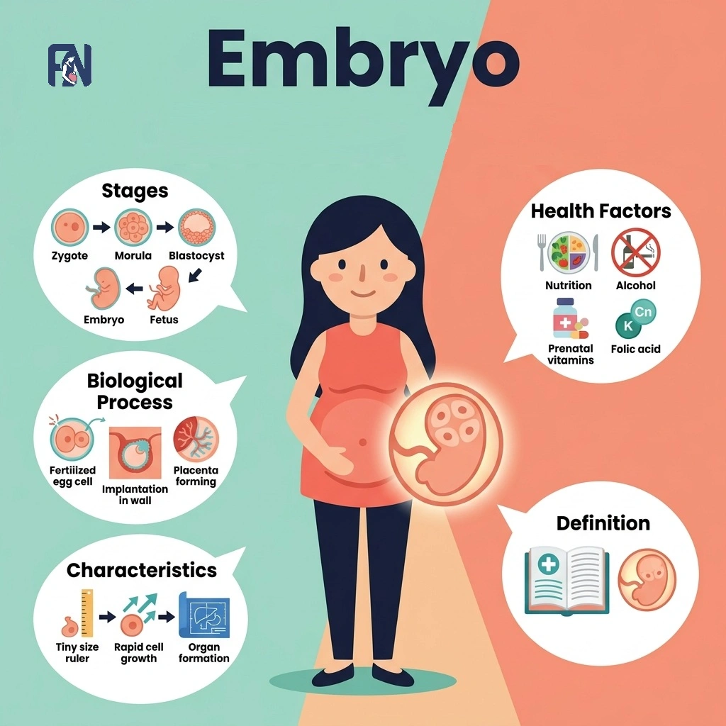
An embryo refers to an early-stage multicellular organism developing from a fertilized egg before major body systems form. The main stages of human embryonic development include zygote, blastocyst, embryo, and fetus, marking critical transitions in prenatal growth. Embryo formation begins biologically with fertilization, the union of sperm and egg, and proceeds through rapid cell division, implantation, and the progressive differentiation of tissues. Important characteristics of an embryo include cellular totipotency, rapid mitotic division, bilateral symmetry, and the early formation of organ rudiments.
The embryo developmental process includes blastocyst formation by day 5, uterine implantation by the end of week 2, and the completion of major organ structures by week 8. The main factors affecting embryo health include maternal age, genetic composition, hormonal balance, nutritional status, and environmental exposures, each of which can contribute to developmental abnormalities when disrupted.
Understanding embryonic health is fundamental to preventing pregnancy complications and improving reproductive outcomes for families affected by infertility. Fertility Network connects patients across India with qualified fertility specialists and evidence-based treatments to support healthy embryonic development at every stage of the process.
What Is an Embryo?
An embryo is a multicellular prenatal organism at its earliest stage of development organism, formed immediately after fertilization of an egg by sperm and developing continuously through the eighth week of gestation.
The difference between an animal embryo and a human embryo is that animal embryos may develop externally within eggs or inside various anatomical structures, whereas a human embryo develops exclusively within the uterus following implantation into the endometrial lining. The embryo forms when fertilization produces a single-celled zygote that undergoes successive mitotic divisions, generating a progressively more complex multicellular structure over the following days and weeks.
The term “embryo” in its modern medical usage was formally established in the 19th century as anatomical researchers began systematically distinguishing prenatal developmental stages and describing the structural sequence from fertilization onward. According to the presentation data from the American Society for Reproductive Medicine (ASRM) showed that incubating eggs in specialized, nutrient-dense media for just 3 to 6 hours before fertilization significantly improved overall embryo development, independent of maternal age.
What is The Location of an Embryo?
The location of an embryo is inside the uterus (womb), where it attaches to the endometrial lining and continues to develop across the different stages of early intrauterine pregnancy. The fertilized egg attaches to the uterine lining, known as the endometrium, after migrating from the fallopian tube, and this attachment marks the true beginning of the embryonic stage proper.
Implantation commonly occurs in the upper portion of the uterus, called the fundus, where a richly vascularized environment supplies the hormonal signals and nutrients that sustain the embryo through the critical early weeks of development.
What is the Duration of the Full Embryonic Development?
The duration of the full embryonic development is from fertilization through the 8 weeks of pregnancy, or approximately 56 days, during which all primary organ systems are established within the developing organism inside the uterine environment.
The embryonic period begins with the process of fertilization and continues until the completion of the eighth week of development, marked by rapid cell division, tissue formation, and the development of major organs and body structures that define the human body plan. Following the embryonic stage, development transitions into the fetal stage, during which all established organ systems grow, mature, and prepare for postnatal independent function outside the womb.
What are the Stages of Human Embryonic Development?
The stages of human embryonic development include fertilization, cleavage, blastocyst formation, implantation, gastrulation, organogenesis, and the fetal stage, progressing from a single-cell zygote to a fully developed baby.
The stages of human embryonic development are listed below.
- Fertilization Stage (12–24 Hours Post-Ovulation): The fertilization stage refers to the biological union of a sperm cell and an oocyte in the ampullary region of the fallopian tube, producing a single diploid zygote that carries the complete and unique chromosomal arrangement of both parents. This newly formed zygote contains all genetic information required to direct every subsequent stage of embryonic development. Fertilization marks the biological origin point of a new human organism and initiates the cascade of cellular events that follow.
- Cleavage Stage (Days 1–4): The cleavage stage refers to a rapid series of mitotic divisions of the zygote that produce progressively smaller daughter cells called blastomeres, without any net increase in the total cellular volume of the developing mass. This process begins approximately 24 hours after fertilization and continues through day 4, transforming the single-celled zygote into a compact, mulberry-shaped multicellular structure known as the morula. The cleavage stage establishes the cellular foundation upon which all subsequent structural differentiation depends.
- Blastocyst Stage (Days 5–7): The blastocyst stage refers to the phase in which the morula undergoes blastulation, transforming into a hollow, fluid-filled structure characterized by an internal cavity called the blastocoel, an outer trophoblast layer, and an inner cell mass of pluripotent cells. The trophoblast layer initiates the biochemical preparation for uterine implantation, while the inner cell mass contains all cells destined to form the embryo itself. This stage concludes the pre-implantation period and positions the developing structure for physical attachment to the maternal endometrium.
- Implantation Stage (Days 6–7 to End of Week 2): The implantation stage refers to the process by which the blastocyst embeds itself into the endometrial lining of the uterus, initiating a physical and biochemical connection between the embryo and the maternal body. This process begins around days 6 to 7 post-fertilization and is typically completed by the end of the second week of development. Successful implantation establishes the nutritional and hormonal exchange essential for all sustained embryonic development that follows.
- Gastrulation Stage (Weeks 2–3): The gastrulation stage refers to the reorganization of the bilaminar embryonic disc into three primary germ layers, ectoderm, mesoderm, and endoderm, occurring between weeks 2 and 3 of development. Each germ layer serves as the developmental source of distinct organ systems, collectively establishing the foundational body plan of the entire organism. Gastrulation transforms a structurally simple disc into a trilaminar embryo with defined axes, polarity, and tissue identity.
- Organogenesis Stage (Weeks 3–8): The organogenesis stage refers to the critical developmental period during which the three primary germ layers differentiate into organ rudiments and specialized tissues, driving the formation of all major body systems including the neural tube, cardiac structures, limb buds, and the digestive, respiratory, and urogenital systems. This stage spans weeks 3 through 8 of the embryonic period and represents the most teratogenically sensitive window of human development, as all primary organ systems are actively forming. Organogenesis concludes with the structural completion of all major organs and marks the biological transition from embryo to fetus.
- Fetal Stage (Week 9 to Birth): The fetal stage refers to the developmental period beginning at week 9 and continuing through delivery, defined by progressive organ maturation, functional refinement, and overall growth rather than the primary formation of new structures. All organ systems established during organogenesis undergo continuous specialization and physiological preparation for extrauterine life throughout this period. The fetal stage represents the longest phase of prenatal development and culminates in a fully formed organism capable of independent survival at birth.
What Is the Difference Between Embryo and Fetus?
The difference between an embryo and a fetus is that an embryo is the early stage, up to 8 weeks post-fertilization, while a fetus is the later stage from the 9th week until birth.
An embryo becomes a fetus at the conclusion of the eighth gestational week, once the foundational architectural organization of the human body has been structurally established and all primary organs have formed. The characteristics of an embryo transition into a fetus as organ formation gives way to organ growth and maturation, with the fetal period marked by increasing body size, the refinement of organ systems, and physiological preparation for independent life outside the womb.
What are The Characteristics of an Embryo?
The characteristics of an embryo include rapid mitotic division, cellular totipotency and pluripotency, bilateral symmetry, organ rudiment formation, developmental plasticity, and dependence on the uterine environment.
The characteristics of an embryo are listed below.
- Rapid Mitotic Division: Rapid mitotic division refers to the accelerated rate of cellular multiplication in which the zygote progresses from a single cell to a complex multicellular organism within days following fertilization. This process generates the necessary cellular mass for tissue specification and subsequent organ formation throughout the embryonic stage.
- Cellular Totipotency and Pluripotency: Cellular totipotency and pluripotency refer to the extraordinary capacity of early embryonic cells to differentiate into any cell type present in the human body, a defining biological characteristic of the earliest stages of embryonic development. Totipotency is characteristic of the initial cleavage-stage cells, while pluripotency becomes the dominant property within the inner cell mass of the blastocyst.
- Bilateral Symmetry: Bilateral symmetry refers to the structural organization of the developing embryo in which a definable left-right axis emerges along the central body plane during gastrulation, establishing the architectural template for the placement of all major organ systems. This feature governs the organized and reproducible arrangement of developing organs within the growing embryonic body.
- Organ Rudiment Formation: Organ rudiment formation refers to the emergence of early structural precursors to the major organ systems, including the neural tube, cardiac loops, and limb buds, during the period from weeks 3 through 8 of embryonic development. These rudiments serve as the biological foundation from which fully differentiated and functionally mature organs will subsequently develop during the fetal period.
- Developmental Plasticity: Developmental plasticity refers to the embryo’s capacity to adjust its developmental trajectory in response to environmental signals, epigenetic regulatory mechanisms, or disruptions in maternal physiology encountered during critical windows of organogenesis. Developmental plasticity affects the embryo by modifying its growth in response to environmental signals. While this flexibility enables adaptive responses, it also creates vulnerability, as external perturbations during sensitive developmental periods can permanently alter normal embryonic growth patterns.
- Dependence on Uterine Environment: Dependence on the uterine environment refers to the embryo’s complete reliance on the endometrium and its associated vascular supply for oxygen, nutrients, and the hormonal signals required to sustain its development during the earliest weeks of pregnancy. Inadequate uterine receptivity, compromised endometrial vascularization, or hormonal insufficiency can impair the embryo’s capacity to implant and develop normally.
What Factors Affect Embryo Health?
The factors that affect an embryo include maternal age, genetic composition, hormonal balance, nutritional status, environmental exposures, and uterine health.
The factors that affect an embryo are listed below.
- Maternal Age: Maternal age refers to the chronological age of the mother at conception, which directly influences the chromosomal integrity of the oocyte and the developmental quality of the embryo produced following fertilization. Advanced maternal age is associated with significantly increased rates of chromosomal abnormalities and reduced embryo viability and implantation success.
- Genetic Composition: Genetic composition refers to the complete inherited genetic material contributed by both parents, which determines the embryo’s developmental blueprint and its susceptibility to inherited disorders or abnormalities in cellular function and tissue organization. Mutations, chromosomal rearrangements, or gene deletions in parental genetic material can be transmitted directly to the embryo and disrupt normal developmental signaling pathways.
- Hormonal Balance: Hormonal balance refers to the precise regulation of reproductive hormones, including estrogen, progesterone, and human chorionic gonadotropin, that collectively orchestrate endometrial preparation, embryo implantation, and the early physiological support of the developing embryo. Disruptions in these hormonal signals can compromise endometrial receptivity and lead to embryo developmental abnormalities, implantation failure, or early pregnancy loss.
- Nutritional Status: Nutritional status refers to the adequacy of the mother’s intake of essential micronutrients, including folate, iron, iodine, and omega-3 fatty acids, that are required to support the biochemical processes underlying embryonic cell division, organogenesis, and tissue development. A clinical study published in Reproductive BioMedicine Online (2026) found that women who adhered to a strict pro-fertility diet combined with daily omega-3 fatty acid supplementation for 60 days significantly increased their fertilization rates. It also led to a higher proportion of top-tier Grade A and B embryos. Nutritional deficiencies during this period are linked to neural tube defects, intrauterine growth restriction, and other developmental abnormalities.
- Environmental Exposures: Environmental exposures refer to teratogenic agents, including alcohol, tobacco, certain medications, ionizing radiation, and industrial chemicals, that can interfere with normal embryonic cellular division and differentiation when encountered during sensitive developmental windows. Even brief or limited exposure to specific teratogens during the embryonic period can produce lasting structural or functional abnormalities in the developing organ systems.
- Uterine Health: Uterine health refers to the structural and functional integrity of the uterus, encompassing the quality of the endometrial lining and the absence of anomalies such as fibroids, polyps, or intrauterine adhesions that may obstruct implantation or impair embryo development. A compromised uterine environment reduces the embryo’s capacity to implant successfully and receive the sustained nourishment required for healthy continued development.
What are The Common Disorders and Developmental Abnormalities of an Embryo?
Common disorders and developmental abnormalities include genetic abnormalities, chromosomal conditions, and developmental defects, each of which can compromise the integrity and trajectory of embryonic development in clinically significant ways.
The common disorders and developmental abnormalities are listed below.
- Genetic Abnormalities: Genetic abnormalities refer to mutations or structural DNA errors within the genomic sequence that disrupt normal gene function and impair the precise regulatory mechanisms governing embryonic cellular differentiation and development. These errors may arise spontaneously during gamete formation or early cell division and can affect the organization, differentiation, and long-term survival of embryonic cell populations.
- Chromosomal Conditions: Chromosomal conditions refer to numerical or structural deviations in the chromosomal complement of embryonic cells that fundamentally disrupt the developmental instructions encoded within the genome. These conditions include aneuploidy (the presence of an abnormal chromosome number) with clinically prominent examples including Down syndrome (trisomy 21) and Turner syndrome (45, X), each producing distinct and measurable developmental impacts.
- Developmental Defects: Developmental defects refer to structural anomalies arising from disruptions in organogenesis that produce congenital malformations affecting one or more body systems. These defects range from minor structural variations to severe abnormalities incompatible with survival, and they represent a leading contributor to infertility, implantation failure, and recurrent pregnancy loss in clinical reproductive medicine.
Can Embryo Disorders Cause Infertility?
Yes, embryo disorders can cause infertility, particularly when they involve chromosomal abnormalities or genetic mutations that prevent the embryo from developing normally or from implanting successfully within the uterine lining. These disorders often interfere with normal embryo development, preventing successful implantation or continued growth, and they represent a primary contributing factor in recurrent pregnancy loss and repeated failed assisted reproductive cycles.
Early treatments, including preimplantation genetic testing (PGT), chromosomal screening of embryos prior to transfer, and advanced embryo culture protocols, are increasingly used in India and globally to identify and address these conditions before transfer, substantially improving the likelihood of a viable and sustained pregnancy.
What are the Best Embryo Disorder Treatments in India?
The best embryo treatments in India include IVF, ICSI, preimplantation genetic testing, blastocyst culture and transfer, time-lapse embryo monitoring, and endometrial receptivity analysis, representing a comprehensive suite of advanced embryo care options available across major Indian cities and states.
The best embryo treatments in India are listed below.
- IVF (In Vitro Fertilization): IVF is a widely practiced assisted reproductive technology offered at fertility centers across Indian cities including Mumbai, Delhi, Bangalore, Chennai, and Hyderabad, in which eggs are retrieved, fertilized in a laboratory setting, and the resulting embryo is transferred to the uterus. This treatment addresses a broad range of embryo-related infertility causes, with pricing of a standard IVF cycle in India typically ranging from ₹1,50,000 to ₹2,50,000.
- ICSI (Intracytoplasmic Sperm Injection): ICSI refers to a specialized fertilization technique available at fertility centers in cities including Hyderabad, Pune, Kolkata, and Ahmedabad, in which a single sperm cell is directly injected into an egg to achieve fertilization in cases where conventional IVF may be insufficient. This procedure is particularly effective for male-factor infertility and is frequently combined with IVF as a complete embryo care protocol.
- PGT (Preimplantation Genetic Testing): PGT refers to the genetic screening of embryos before uterine transfer, offered at advanced reproductive medicine centers in Delhi, Mumbai, Chennai, and Bengaluru, enabling clinicians to identify and select chromosomally normal embryos for implantation. This Indian treatment significantly reduces the risk of chromosomally driven implantation failure and miscarriage, with pricing of PGT typically ranging from ₹30,000 to ₹60,000 as an add-on to the IVF cycle.
- Blastocyst Culture and Transfer: Blastocyst culture and transfer refers to the extended laboratory cultivation of embryos to day 5 or day 6 before uterine transfer, practiced at fertility centers across states including Maharashtra, Karnataka, Tamil Nadu, and Telangana. Extended culture improves embryo selection accuracy, increases implantation rates, and enhances overall embryo care outcomes compared to earlier-stage transfer protocols.
- Time-Lapse Embryo Monitoring: Time-lapse embryo monitoring refers to a non-invasive imaging system that continuously photographs developing embryos within a specialized incubator, enabling embryologists to assess developmental kinetics and select the most viable embryos without disturbing the culture environment. This technology is available at select premium fertility centers in cities such as Bangalore, Delhi, and Hyderabad, typically included within advanced IVF packages with pricing varying by clinic.
- Endometrial Receptivity Analysis (ERA): ERA refers to a diagnostic procedure in which a uterine biopsy sample is analyzed to identify the precise implantation window for an individual patient, enabling personalized embryo transfer timing that optimizes implantation success. This test is offered across reproductive medicine units in major Indian cities and states, with pricing typically ranging from ₹15,000 to ₹30,000 as part of a comprehensive embryo care cycle.
How Can Modern Indian Treatments Help with Embryo-Related Infertility?
Modern Indian treatments can help address embryo-related infertility by improving embryo quality, supporting uterine receptivity, and increasing the chances of successful implantation through a combination of established and emerging reproductive technologies. Assisted reproductive technologies available at fertility centers across India include IVF, ICSI, IUI, and PGT, each designed to optimize specific aspects of fertilization, embryo development, and implantation success based on the patient’s clinical profile.
All treatments are offered through a different pricing model structured to accommodate a wide range of financial needs and treatment complexities. Emerging supportive therapies such as PRP endometrial rejuvenation, AI-assisted embryo selection, and time-lapse imaging further enhance treatment precision, enabling Indian fertility specialists to intervene at each stage of embryo development using targeted, evidence-based approaches that improve outcomes across the full spectrum of embryo-related infertility presentations.
How Much Does Embryo-Related Infertility Treatment Cost in India?
Embryo-related fertility treatment in India costs between ₹1,50,000 and over ₹4,00,000 per cycle, depending on the clinic, protocol, and medication requirements, making India one of the most accessible and cost-effective destinations globally for high-quality assisted reproductive care. The cost typically includes IVF or ICSI procedures, embryo culture, embryo grading, the stages of embryonic development being addressed during the protocol, embryo transfer, and fertility medications, with specific inclusions varying according to each patient’s diagnosis, the biological process involved, and the fertility center’s standard protocol.
Costs vary further based on city location, the specific evidence-based treatment protocols applied to optimize factors regarding embryo health, embryo grading and selection requirements, and the dosage and duration of fertility medications required to address each patient’s underlying condition. Additional procedures such as PGT, ERA, time-lapse monitoring, and PRP endometrial therapy, along with assessments of the characteristics of an embryo required prior to transfer, may increase the total cost beyond the base cycle fee. Fertility Network can help patients find proper and affordable treatment centers and doctors near them, ensuring access to quality care that appropriately addresses the specific factors that affect embryo health and the characteristics of each patient’s clinical situation.

Fathima Zahra is a reproductive health content specialist at FertilityNetwork.in — India’s largest independent fertility clinic directory and patient education platform.
Fathima’s work covers the full spectrum of reproductive medicine, from foundational biology to advanced fertility treatment. Her published articles span reproductive anatomy (ovaries, uterus, fallopian tubes, testes), core reproductive processes (ovulation, fertilization, implantation, the menstrual cycle), reproductive hormones (estrogen, progesterone, testosterone, FSH, LH, AMH, and prolactin), and the clinical conditions that cause infertility in both men and women.
On the male side, her research covers conditions including azoospermia, oligospermia, asthenozoospermia, teratozoospermia, varicocele, erectile dysfunction, Klinefelter syndrome, and Y chromosome microdeletion. On the female side, she writes about PCOS, endometriosis, uterine fibroids, anovulation, diminished ovarian reserve, premature ovarian insufficiency, tubal blockage, pelvic inflammatory disease, and Turner syndrome.
Her treatment coverage includes intrauterine insemination (IUI), in vitro fertilization (IVF), intracytoplasmic sperm injection (ICSI), egg retrieval, embryo transfer, egg freezing, sperm freezing, and oncofertility — providing patients with detailed, process-level understanding of each procedure, its clinical indications, success rates, and costs.
Every article Fathima publishes follows a structured, evidence-based research methodology:
1. Source verification against ICMR (Indian Council of Medical Research) national fertility and ART guidelines
2. Cross-referencing with ESHRE (European Society of Human Reproduction and Embryology) clinical protocols
3. Alignment with ASRM (American Society for Reproductive Medicine) evidence-based practice guidelines
4. Citation of PubMed-indexed, peer-reviewed studies published within the last 5 years
5. Verification of all diagnostic thresholds, hormonal reference ranges, and clinical statistics against current international standards
All content authored by Fathima undergoes editorial review by the FertilityNetwork Medical Research Board before publication, ensuring clinical accuracy and factual integrity across every article published on the platform.
Fathima’s content is editorially independent. FertilityNetwork.in does not publish sponsored medical opinions, and no article is written to promote specific clinics, doctors, or fertility treatments.
Leave a Reply