Fathima

  • Ovaries: Definition, Location, Anatomy, Function, Phases and Conditions

    Ovaries: Definition, Location, Anatomy, Function, Phases and Conditions

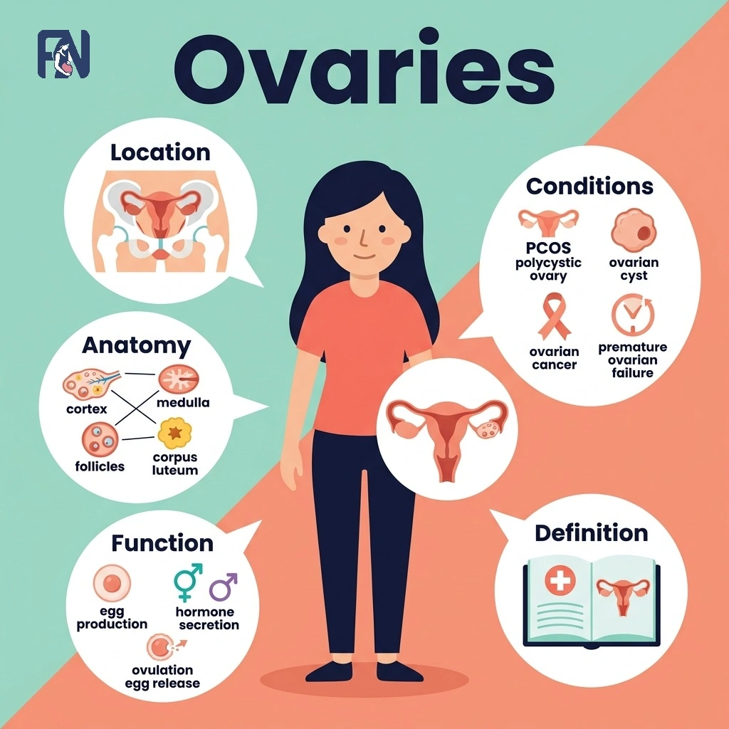
    The ovaries basically refer to the two reproductive organs that store eggs and produce hormones. Women typically have two ovaries, one on each side of the uterus, connected to the fallopian tubes. Each ovary contains follicles that hold immature eggs, along with the tissue and blood vessels that support hormone production. The ovaries produce estrogen and progesterone, which regulate the menstrual cycle, support pregnancy, and influence fertility from puberty through menopause.

    The ovarian cycle consists of the follicular, ovulation, and luteal phases that repeat roughly every month. Some of the major ovarian conditions are PCOS, ovarian cysts, ovarian torsion, premature ovarian insufficiency, and ovarian cancer, along with the symptoms that signal a problem. Regular monitoring of ovarian health through pelvic exams, ultrasounds, and hormone testing helps detect these conditions early, and a doctor should be consulted if symptoms such as pelvic pain or irregular periods persist. Treatment options range from medication and lifestyle changes to laparoscopic surgery, depending on the condition and its severity.

    What Are the Ovaries?

    The ovaries are paired reproductive organs that function as both gonads and endocrine organs, producing female gametes called oocytes while generating the hormones that regulate reproductive function. As gonads, the ovaries sit at the center of the female reproductive system, working alongside the uterus and fallopian tubes to support conception. They carry out the dual role of releasing mature eggs during ovulation and secreting estrogen and progesterone to regulate the menstrual cycle. 

    Most women have two ovaries, which provides a biological backup, since ovulation can continue even if one ovary is removed or damaged. Healthy ovaries are essential for fertility, hormonal balance, and long-term reproductive health. According to recent research by the World Health Organization, infertility impacts roughly 1 in 6 adults globally, with a declining ovarian reserve and poor egg quality serving as leading causes of female infertility.

    Where Are the Ovaries Located?

    The ovaries are located in the pelvis, one on each side of the uterus, below the fallopian tubes and close to the pelvic sidewall. Each ovary sits just below the fimbriated end of its corresponding fallopian tube, held in place by supporting ligaments that connect it to the uterus and pelvic wall. A typical ovary is almond-shaped and measures about 3 to 5 centimeters in length, though its size can shift slightly with the menstrual cycle and age. As with other parts of the anatomy of a woman’s body, exact ovarian position can vary slightly from person to person and may shift with pregnancy or prior pelvic surgery.

    What Is the Anatomy of the Ovaries?

    The anatomy of the ovaries includes the outer cortex, inner medulla, surface epithelium, follicles at various stages of development, and the blood vessels and ligaments that support each ovary. These structures work together to store and mature eggs, produce hormones, and maintain a stable blood supply throughout the menstrual cycle. 

    The main structures of the ovarian anatomy are listed below.

    • Ovarian Structure: The ovarian structure consists of an outer cortex, which contains developing follicles, and an inner medulla containing blood vessels and connective tissue. A layer of germinal epithelium covers the ovary’s surface, protecting its internal structures. Connective tissue and a dedicated blood supply support the cortex and medulla throughout each phase of the reproductive cycle.
    • Ovarian Follicles: Ovarian follicles are small, fluid-filled sacs within the ovary that each contain a single developing egg. Follicles progress through several stages, from primordial and primary follicles to secondary follicles and finally the mature Graafian follicle. This process, known as folliculogenesis, allows one dominant follicle to mature and release its egg during ovulation each cycle.
    • Blood Supply and Ligaments: The blood supply and ligaments of the ovaries include the ovarian artery and ovarian vein, which deliver oxygenated blood and drain the ovary, along with lymphatic drainage that supports immune function. The ovarian ligament anchors the ovary to the uterus, while the suspensory ligament connects it to the pelvic wall and carries the main ovarian blood vessels. Together, these ligaments stabilize the ovary and maintain the vascular supply it needs to function.
    • Oocytes and Egg Development: Oocytes and egg development are the process by which immature egg cells, or oocytes, mature within ovarian follicles through oogenesis, a process tightly regulated by hormone signals. A woman’s full supply of oocytes forms before birth, and this ovarian reserve steadily declines with age, affecting both fertility and hormone production over time. During each menstrual cycle, one oocyte typically completes egg maturation and is released at ovulation in response to a surge of luteinizing hormone, while the rest remain dormant or are lost naturally.

    What Hormones Are Produced by the Ovaries?

    Hormones produced by the ovaries include estrogen, progesterone, testosterone, inhibin, and relaxin, which regulate the menstrual cycle, support fertility, maintain pregnancy, and contribute to overall reproductive health.

    The hormones produced by the ovaries are listed below.

    • Estrogen: Estrogen refers to the primary female sex hormone responsible for puberty, menstrual regulation, and the development of secondary sexual characteristics. Estradiol, the main form of estrogen, supports breast development and helps maintain bone health throughout a woman’s reproductive years. Estrogen levels rise and fall predictably across the menstrual cycle, driving follicle growth and preparing the uterine lining.
    • Progesterone: Progesterone refers to the hormone released mainly by the corpus luteum after ovulation to prepare the endometrium for pregnancy. It thickens and stabilizes the uterine lining to support implantation if fertilization occurs. If pregnancy does not happen, progesterone levels drop, triggering the start of a new menstrual cycle.
    • Testosterone: Testosterone is an androgen produced in small amounts by the ovaries, contributing to libido, muscle tone, and overall hormonal balance in women. It is considered a secondary ovarian hormone. It stimulates sex drive and helps maintain muscle mass and bone density by acting on cells throughout the body. Inhibin is a protein-based secondary hormonal substance released by the ovaries
    • Inhibin: Inhibin is a hormone produced by ovarian follicles that suppresses follicle-stimulating hormone release from the pituitary gland, helping regulate follicle development.
    • Relaxin: Relaxin is a hormone released mainly during the luteal phase and pregnancy that relaxes the uterine muscles and pelvic ligaments to support implantation and later childbirth. Relaxin is a peptide secondary hormonal substance secreted by the ovaries.

    What Is the Hormonal Regulation of the Ovaries?

    Hormonal regulation of the ovaries refers to the feedback loop known as the hypothalamic-pituitary-ovarian axis, which coordinates ovarian functions with signals from the brain. The hypothalamus releases hormones that prompt the pituitary gland to secrete gonadotropins, namely FSH and LH, which stimulate follicle growth and trigger ovulation. In turn, rising and falling estrogen and progesterone levels send hormonal feedback back to the brain, adjusting FSH and LH output throughout the cycle.

    What Are the Functions of the Ovaries?

    The functions of the ovaries include producing and releasing eggs, generating reproductive hormones, and supporting the physical processes that make conception and pregnancy possible. 

    The main functions of the ovaries are listed below.

    • Egg Production: Egg production refers to the ovaries’ ongoing process of developing ova each month through oocyte maturation within growing follicles. Multiple follicles begin developing at the start of each reproductive cycle, though typically only one reaches full maturity. That dominant follicle releases its egg during ovulation, making it available for fertilization.
    • Ovulation: Ovulation refers to the release of a mature egg from the dominant follicle, typically occurring around the midpoint of the menstrual cycle. A surge in luteinizing hormone (LH) triggers follicular rupture, allowing the egg to travel into the fallopian tube. This event marks the beginning of the fertile window, when conception is most likely.
    • Hormone Production: Hormone production in the ovaries refers to their endocrine function of secreting estrogen and progesterone, the primary female sex hormones. These hormones regulate the menstrual cycle, support pregnancy, and maintain hormonal balance throughout a woman’s reproductive years. Disruptions in ovarian hormone production can affect fertility, bone health, and overall well-being.
    • Reproductive Function: The reproductive function of the ovaries refers to their role in enabling fertilization, implantation, and pregnancy by working together with the uterus and fallopian tubes. The ovaries supply the egg, while the fallopian tubes provide the site for fertilization and the uterus supports implantation. This coordinated system underlies female fertility from ovulation through early pregnancy.

    What Is the Ovarian Cycle?

    The ovarian cycle is the monthly hormonal process that governs egg development, and it consists of 3 phases, including the follicular phase, ovulation, and the luteal phase. 

    The 3 phases of the ovarian cycle are listed below.

    • Follicular Phase (Days 1–13): The follicular phase refers to the first part of the ovarian cycle, beginning on the first day of menstruation and lasting until ovulation. This phase typically spans from day 1 to day 13 of a standard 28‑day cycle. Rising estrogen production drives follicle growth, allowing several follicles to develop before one becomes dominant. This phase also thickens the uterine lining in preparation for a potential pregnancy.
    • Ovulation (Day 14): The ovulation phase refers to the brief midcycle period when a surge in luteinizing hormone triggers the release of a mature egg. This phase typically occurs on day 14 of a standard 28‑day cycle. This LH surge causes the dominant follicle to rupture, releasing the egg on ovulation day. The released egg then travels into the fallopian tube, where fertilization can occur if sperm are present.
    • Luteal Phase (Days 15–28): The luteal phase refers to the final stage of the ovarian cycle, occurring after ovulation and lasting about two weeks. This phase spans from day 15 to day 28 of the cycle. During this phase, the corpus luteum produces progesterone, supporting endometrial preparation for possible implantation. If fertilization does not occur, progesterone levels fall, the corpus luteum breaks down, and menstruation begins, restarting the cycle relevant to fertility.

    What Is the Connection Between Ovaries and Fertility?

    The connection between ovaries and fertility is direct, since the ovaries store, mature, and release the eggs needed for conception, and they produce hormones that regulate ovulation and support early pregnancy. Each month, the ovaries prepare and release an egg during ovulation, making fertilization possible if it meets sperm in the fallopian tube. Problems with egg production, ovulation, or hormone balance can make it more difficult to conceive, which is why healthy ovarian function is closely tied to overall fertility.

    How to Keep Ovaries Healthy?

    To keep ovaries healthy, follow the tips mentioned below.

    • Maintain a Healthy Weight: A healthy weight is a body weight within a normal range that supports balanced hormone levels and regular ovulation, since excess or very low body fat can disrupt reproductive health.
    • Exercise Regularly: Exercise is regular physical activity that improves blood flow, supports hormonal balance, and helps maintain a healthy weight, all of which benefit ovarian function.
    • Eat a Balanced Diet: A balanced diet is an eating pattern rich in whole foods, healthy fats, and antioxidants that supports egg quality and hormone production over time.
    • Practice Preventive Care: Preventive care is a proactive approach to reproductive health, including avoiding smoking and managing stress, that helps protect long-term ovarian function.
    • Schedule Regular Checkups: Regular checkups are periodic visits with a gynecologist that allow early detection of ovarian conditions through exams, ultrasounds, or hormone testing.

    Is It Possible to Get Pregnant With One Ovary?

    Yes, it is possible to get pregnant with one ovary because the remaining ovary can ovulate every cycle, maintaining normal fertility in most cases. Even after surgery or an ovarian condition that requires removing one ovary, the other typically compensates by releasing an egg monthly. Fertility may be somewhat reduced only if the remaining ovary also has an underlying issue.

    What Are the Common Ovarian Conditions?

    The common ovarian conditions include PCOS, ovarian cysts, ovarian torsion, premature ovarian insufficiency, and ovarian cancer

    The common ovarian conditions are listed below.

    • Polycystic Ovary Syndrome (PCOS): PCOS refers to a hormonal disorder in which the ovaries produce excess androgens and develop multiple small follicles, disrupting normal ovulation and menstrual function. Hormonal imbalance and insulin resistance increase the risk of PCOS and commonly cause irregular periods, acne, and excess hair growth. Untreated PCOS may lead to infertility and increase the risk of metabolic disorders.
    • Ovarian Cysts: Ovarian cysts refer to fluid-filled sacs that develop on or within an ovary and vary in size, type, and potential health effects. They may occur as a functional cyst, dermoid cyst, or hemorrhagic cyst and can cause pelvic pain, bloating, or pressure. If left untreated, some cysts may rupture, bleed, or lead to ovarian torsion.
    • Ovarian Torsion: Ovarian torsion is the twisting of an ovary around its supporting tissues, reducing or completely blocking its blood supply. A twisted ovary is usually associated with an ovarian cyst or enlarged ovary and is considered an emergency condition. Symptoms include sudden severe pelvic pain, and delayed treatment can result in loss of the ovary.
    • Premature Ovarian Insufficiency: Premature ovarian insufficiency refers to a condition in which the ovaries lose normal function before the age of forty because of early ovarian failure. It causes hormone deficiency, irregular or absent periods, and symptoms of early menopause, including hot flushes and vaginal dryness. The condition can lead to infertility and increase the risk of osteoporosis.
    • Ovarian Cancer: Ovarian cancer is the uncontrolled growth of abnormal cells in the ovary that form an ovarian tumor and may spread to other tissues. Risk factors include age, inherited genetic mutations, and family history, while cancer symptoms may include bloating, pelvic pain, and changes in appetite, making early detection important. Diagnosis may involve imaging and a CA-125 blood test, and treatment can include surgery and chemotherapy to prevent serious complications.

    What Are the Symptoms of Ovarian Problems?

    The symptoms of ovarian problems include pelvic pain, bloating, irregular periods, abdominal swelling, infertility, and pain during intercourse. 

    The symptoms of ovarian problems are listed below.

    • Pelvic Pain: Pelvic pain refers to discomfort in the lower abdomen that can range from mild to severe. It can result from ovarian cysts, inflammation, or other ovarian conditions affecting the surrounding tissue.
    • Bloating: Bloating refers to the feeling of fullness or swelling in the abdomen. It may result from fluid buildup or an enlarged ovary pressing on nearby organs.
    • Irregular Periods: Irregular periods refer to menstrual cycles that vary in length, timing, or flow from month to month. Hormonal imbalance caused by ovarian conditions such as PCOS can disrupt cycle regularity.
    • Abdominal Swelling: Abdominal swelling refers to visible or noticeable enlargement of the abdomen. It can indicate an enlarged ovary, an ovarian cyst, or, in rare cases, ovarian cancer.
    • Infertility: Infertility refers to the inability to conceive after a year or more of regular, unprotected intercourse. Disrupted ovulation or egg quality issues linked to ovarian conditions can prevent conception.
    • Pain During Intercourse: Pain during intercourse refers to discomfort or sharp pain felt during or after sexual activity. It can result from ovarian cysts, endometriosis, or pelvic inflammation affecting nearby structures.

    For any of the symptoms above ealy diagnosis playes a vital role in a woman’s life.

    How Do Doctors Diagnose Ovarian Health?

    Doctors evaluate ovarian health using a combination of physical examination, imaging, and laboratory testing to check for cysts, hormonal imbalances, and other abnormalities. 

    The common methods used to evaluate ovarian health are listed below.

    • Pelvic Examination: A pelvic examination is a physical exam in which a doctor checks the ovaries, uterus, and surrounding structures for abnormalities. It helps doctors detect tenderness, masses, or irregularities and is typically recommended during routine gynecological visits or when symptoms arise.
    • Ultrasound: An ultrasound, including a transvaginal ultrasound, is an imaging test that uses sound waves to create detailed images of the ovaries. It helps visualize the ovaries and detect cysts, tumors, or other structural abnormalities.
    • Hormone Testing: Hormone testing involves blood tests that measure reproductive hormone levels to assess ovarian function. Common markers include AMH, FSH, and estradiol, which help evaluate ovarian reserve and hormonal balance.
    • Fertility Testing: Fertility testing involves a series of exams and blood tests that evaluate ovarian reserve and function. Results help guide fertility treatment decisions, including whether medication, IUI, or IVF may be appropriate.

    What Are the Best Treatments for Ovarian Conditions in India?

    The best treatments for ovarian conditions in India include medication, minimally invasive surgery, and advanced fertility care available across major metropolitan cities with experienced specialists and modern diagnostic facilities. The best treatments for ovarian conditions in India are listed below.

    • Delhi: Delhi offers advanced gynecological and fertility centers with specialists in PCOS management, laparoscopic surgery, and IVF, with treatment costs varying by procedure and hospital.
    • Mumbai: Mumbai is home to leading fertility hospitals offering ovarian cyst removal, hormone therapy, and IVF, with a wide cost range depending on treatment complexity.
    • Bangalore: Bangalore provides access to reproductive endocrinologists and fertility clinics specializing in ovarian condition diagnosis and treatment, with costs varying by clinic and procedure type.
    • Chennai: Chennai has established fertility centers offering ovarian surgery, hormone testing, and IVF services, with treatment cost ranges depending on the specific ovarian condition.
    • Hyderabad: Hyderabad offers well-equipped fertility hospitals providing laparoscopic ovarian surgery and hormonal treatment, with costs varying based on diagnosis and required procedures.
    • Kolkata: Kolkata has growing fertility infrastructure with specialists treating PCOS, ovarian cysts, and infertility, offering a range of cost options depending on treatment type.
    • Pune: Pune provides access to fertility specialists and diagnostic centers for ovarian conditions, with treatment cost ranges depending on the chosen hospital and procedure.

    How Much Does Ovarian Condition Treatment Cost in India?

    The cost of ovarian condition treatment in India ranges from ₹5,000 to ₹10,00,000 depending on the diagnosis, treatment approach, and hospital chosen. Basic medication and hormone therapy sit at the lower end, while laparoscopic procedures cost more due to surgical expertise and facility charges. Complex surgeries, such as those for ovarian cancer or advanced endometriosis, fall at the higher end of the cost range.

    How Do Ovarian Conditions Cause Infertility?

    Ovarian conditions can cause infertility via disruption of ovulation, reduced egg quality, or hormonal imbalance needed for successful conception. Conditions such as PCOS, premature ovarian insufficiency, endometriosis, and ovarian cysts can directly affect reproductive health, decrease fertility, and lead to infertility. Early diagnosis and proper treatment may improve reproductive outcomes and increase the chances of pregnancy.

    How to Find Proper Treatments in India for Infertility Caused by Ovarian Problems?

    To find proper treatments in India for infertility caused by ovarian conditions, consult a fertility specialist for an accurate diagnosis and personalised care. The assessment should examine the location of ovaries, their anatomy, and the functions of ovaries to understand their role in fertility. Doctors should also assess the different phases of the menstrual cycle and determine how specific ovarian conditions affect fertility. It is important to choose clinics that provide evidence-based treatment and have experience managing infertility related to ovarian disorders. Fertility Network helps people find affordable fertility treatment options near them in India and connect with qualified fertility specialists.
  • Testicles (Testes): Definition, Location, Anatomy, Function and Conditions

    Testicles (Testes): Definition, Location, Anatomy, Function and Conditions

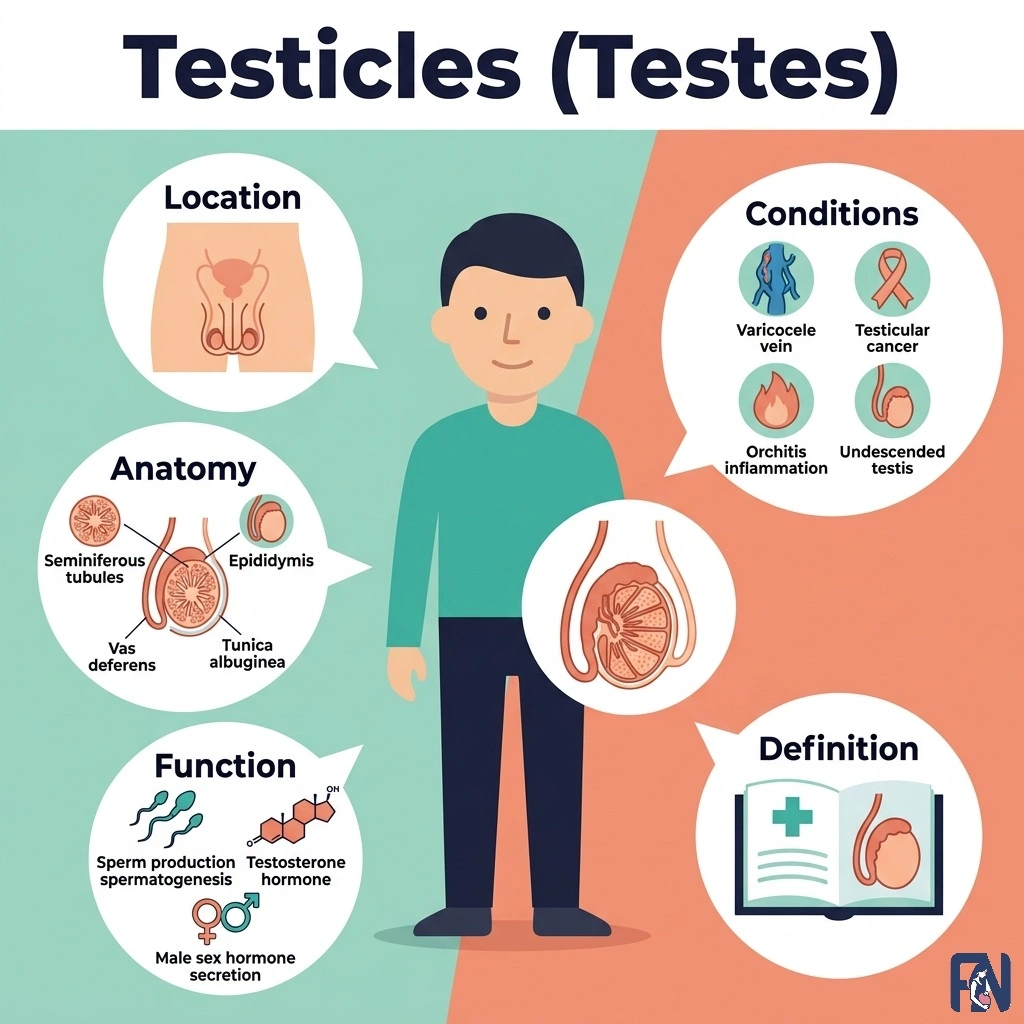
    The testicles refer to the two male reproductive glands that produce sperm and testosterone. Testes are the primary reproductive organs of the male reproductive system. They are located in the scrotum and work continuously to support fertility and hormone production. Testosterone from the testicles drives sperm production and also shapes muscle mass, bone density, and overall male health. Understanding testicular anatomy and function makes it easier to recognize when something feels different and to seek care early.

    Some common testicular conditions include testicular cancer, testicular torsion, epididymitis, orchitis, varicocele, and hydrocele. Monitoring testicular health through regular self-exams and medical diagnosis of new lumps, swelling, or pain helps catch problems while they are most treatable. Treatment options range from antibiotics and lifestyle changes to surgery, orchiectomy, or chemotherapy, depending on the condition and its severity.

    What Are Testicles?

    Testicles, also known as testes, are the male gonads located within the scrotum, working continuously to produce sperm and testosterone for reproduction. There are two of these glands, each situated on opposite sides of the scrotum, functioning independently to maintain male fertility and hormonal balance. Testes work by using specialized cells inside each testicle to generate sperm and release hormones directly into the bloodstream, keeping the male reproductive system running on a steady cycle. 

    Maintaining good testicular health is essential, as any injury, infection, or hormonal imbalance affecting these glands can disrupt sperm production, reduce testosterone levels, and impact overall male fertility and well-being. A 2022 editorial in Frontiers in Endocrinology indicates that male factors contribute to nearly 50% of failed pregnancy attempts, with a 20% rate as the sole cause and a 30% rate as a contributing factor.

    Where Are Testicles Located?

    Testicles are located in the scrotum, a pouch of skin that hangs outside the body just below the penis. This external position keeps the testicles about 2 to 4 degrees cooler than the rest of the body, which is essential for healthy sperm production, since sperm cannot develop properly at core body temperature. A typical adult testicle measures about 4 to 5 centimeters in length, roughly the size of a large olive. Understanding this part of male anatomy helps explain why conditions like excessive heat exposure can affect fertility. 

    What Is the Anatomy of the Testicles?

    The anatomy of the testicles includes the seminiferous tubules, epididymis, spermatic cord, and the Leydig and Sertoli cells that support sperm and hormone production. Each structure plays a distinct role in producing, transporting, or regulating sperm and testosterone. 

    The main structures of the testicular anatomy are listed below.

    • Seminiferous Tubules: The seminiferous tubules are the tightly coiled structures inside each testicle where sperm cells are produced. They make up most of the testicle’s internal volume and are lined with specialized cells that support sperm development. This is the primary site of sperm production within the testicles.
    • Epididymis: The epididymis refers to the coiled tube attached to the back of each testicle that connects it to the vas deferens. Sperm travel through the epididymis after leaving the seminiferous tubules, gaining the ability to swim and fertilize an egg. It stores and matures sperm before ejaculation, typically over a period of about two weeks.
    • Spermatic Cord: The spermatic cord refers to the bundle of tissue that suspends each testicle within the scrotum and connects it to the rest of the body. It contains blood vessels, nerves, and the vas deferens, which together supply and support each testicle. Damage or twisting of the spermatic cord can disrupt blood flow and testicular function.
    • Leydig and Sertoli Cells: Leydig and Sertoli cells are the two main types of specialized cells found within testicular tissue. Leydig cells produce testosterone in response to hormone signals from the brain, directly influencing hormone levels throughout the body. Sertoli cells support developing sperm cells as they mature within the seminiferous tubules.

    What Hormones Are Released by the Testes?

    The hormones released by the testes are listed below.

    • Testosterone: Testosterone is the primary male sex hormone produced by Leydig cells within the testicles. It drives sperm production, sex drive, and the development of secondary male characteristics such as facial hair and a deeper voice. Testosterone levels are tightly regulated by signals from the brain to maintain steady function.
    • Inhibin B: Inhibin B is a hormone produced by Sertoli cells within the testicles that directly reflects the health of ongoing sperm production.Testosterone and inhibin B production are governed by the hypothalamic-pituitary-gonadal (HPG) axis, through which the brain releases LH and FSH to stimulate the testes, while the testes send hormonal feedback to maintain balanced function. Low inhibin B levels can indicate reduced testicular function.
    • Anti-Müllerian Hormone (AMH): AMH is a hormone produced by Sertoli cells within the testicles, essential during fetal development for suppressing female reproductive structures and ensuring proper male tract formation. In adulthood, it continues to be released and serves as a valuable clinical marker of Sertoli cell health and sperm-producing capacity.
    • Estradiol: Estradiol is a form of estrogen produced in small amounts by the testes, primarily through the conversion of testosterone within Sertoli and Leydig cells. It helps modulate libido, supports bone density, regulates brain function, and aids in sperm maturation. Balanced estradiol levels are essential, as extremes can disrupt fertility and overall well-being.
    • Insulin-like factor 3 (INSL3): INSL3 is a hormone produced almost exclusively by Leyd ig cells within the testicles, playing a crucial role during fetal development by guiding testicular descent into the scrotum. In adulthood, it serves as a reliable marker of Leydig cell function and contributes to bone metabolism, muscle health, and reproductive tissue maintenance.

    What Are the Functions of the Testicles?

    The functions of the testicles include producing sperm, generating testosterone, and regulating temperature to support healthy sperm development. 

    The main functions of the testicles are listed below.

    • Sperm Production: Sperm production refers to the testicles’ ongoing process of generating new sperm cells within the seminiferous tubules. This process, called spermatogenesis, occurs continuously throughout adult life and takes about 64 to 72 days to complete. Consistent sperm production is essential for maintaining male fertility.
    • Testosterone Production: Testosterone production refers to the testicles’ role in synthesizing and releasing testosterone, the primary male hormone. This process supports male hormone balance and drives the development of secondary sexual characteristics, including muscle mass and body hair. Testosterone levels typically peak in early adulthood and gradually decline with age.
    • Other Hormonal Regulation: Other hormonal regulation means the testicles’ production of additional hormones beyond testosterone, primarily through Sertoli and Leydig cells. These hormones include Inhibin B, Anti-Müllerian Hormone (AMH), estradiol, and insulin-like growth factor 3 (INSL3), each playing distinct roles in fertility and development. For instance, Inhibin B and AMH help regulate sperm production and fetal reproductive tract formation, while estradiol and INSL3 support bone health, libido, and testicular descent. 
    • Temperature Regulation: Temperature regulation is the scrotum’s ability to keep the testicles slightly cooler than the rest of the body. This cooler environment supports healthy sperm production, since sperm development is highly sensitive to heat. Prolonged heat exposure from conditions such as tight clothing or frequent hot baths can reduce sperm quality over time.

    What Are the Common Testicular Conditions?

    The common testicular conditions include testicular cancer, testicular torsion, epididymitis, orchitis, varicocele, and hydrocele.

    The common testicular conditions are listed below.

    • Testicular Cancer: Testicular cancer refers to a malignant growth that develops within the tissue of a testicle, most often affecting men between the ages of 15 and 35. Risk factors include an undescended testicle, family history, and prior testicular cancer, though the exact cause is often unclear. Common symptoms include a painless lump or swelling, and possible complications include the cancer spreading to nearby lymph nodes or organs if untreated.
    • Testicular Torsion: Testicular torsion is a twisted spermatic cord that cuts off blood supply to the testicle. It is considered an emergency condition, often causing sudden and severe pain along with swelling and nausea. Without prompt treatment, testicular torsion can lead to permanent loss of the affected testicle.
    • Epididymitis: Epididymitis is the inflammation of the epididymis, most often caused by a bacterial infection, including sexually transmitted infections. Symptoms typically include pain, swelling, and tenderness on one side of the scrotum, sometimes with fever. Left untreated, epididymitis can lead to abscess formation or reduced fertility over time.
    • Orchitis: Orchitis refers to inflammation of one or both testicles, commonly caused by a viral or bacterial infection, including the mumps virus. Symptoms include swelling, tenderness, and a feeling of heaviness in the affected testicle. Possible complications include reduced testicular size and, in some cases, impaired fertility.
    • Varicocele: A varicocele refers to an enlargement of the veins within the scrotum, similar to varicose veins elsewhere in the body. It commonly develops during puberty and may cause a dull ache or heaviness, especially after standing for long periods. Left unmanaged, a varicocele can contribute to reduced sperm quality and infertility.
    • Hydrocele: A hydrocele refers to a fluid buildup around a testicle that causes the scrotum to swell. It often develops without a clear cause in adults, while in infants it may result from incomplete closure of a natural passage. Symptoms are usually limited to painless swelling, though large hydroceles can cause discomfort or a feeling of heaviness.

    What Are the Symptoms of Testicular Problems?

    The common symptoms of testicular problems are listed below. 

    • Lump: A testicular lump refers to a firm or hard area felt on or within the testicle. It can be a sign of testicular cancer and requires prompt medical evaluation.
    • Swelling: Testicular swelling refers to visible enlargement of one or both testicles. It may result from infection, fluid buildup, or a varicocele affecting the surrounding tissue.
    • Pain: Testicular pain refers to discomfort ranging from mild aching to sharp, severe pain in one or both testicles. Sudden, severe pain may signal testicular torsion, an emergency condition requiring immediate care.
    • Heaviness: Testicular heaviness refers to a dragging or weighted sensation in the scrotum. This condition is commonly associated with a varicocele or hydrocele affecting one of the testicles.

    How Are Testicular Conditions Diagnosed?

    The methods used to diagnose testicular conditions are listed below.

    • Physical Examination: A physical examination is a hands-on exam in which a doctor checks the size, shape, and texture of each testicle. Doctors look for lumps, swelling, tenderness, or asymmetry that might indicate an underlying condition.
    • Ultrasound: An ultrasound is an imaging test that uses sound waves to create detailed pictures of the testicles. It helps visualize the testicles and detect abnormalities such as tumors, cysts, or torsion.
    • Tumor Markers: Tumor markers are substances measured through blood tests that can indicate the presence of testicular cancer. Alongside a testicular examination and ultrasound, tumor marker testing helps confirm a diagnosis and guide treatment planning.

    What Is a Testicular Self-Examination?

    A testicular self-examination is a simple screening check performed at home to feel for lumps, swelling, or changes in the testicles. Most doctors recommend performing a self-exam once a month, ideally after a warm shower when the scrotal skin is relaxed. Healthy testes typically feel smooth, firm, and slightly rubbery, without hard lumps or sudden changes in size. This routine supports early detection of testicular cancer and other conditions. Any new lump, persistent pain, or noticeable change in size or shape should prompt a visit to a doctor.

    How to Keep Testicles Healthy?

    The habits that support testicular health are listed below.

    • Limit Heat Exposure: Limiting heat exposure means avoiding prolonged use of hot tubs, saunas, and tight clothing, since excess heat can impair sperm production over time.
    • Wear Protective Equipment: Wearing protective equipment, such as an athletic cup during contact sports, helps prevent injury that could damage testicular tissue or function.
    • Maintain a Healthy Lifestyle: Maintaining a healthy lifestyle, including regular exercise and a balanced diet, supports hormone balance and long-term testicular health.
    • Seek Quick Treatment: Seeking quick treatment for pain, swelling, or lumps ensures conditions are caught early, when treatment tends to be simpler and more effective.

    What Are the Best Treatments for Testicular Conditions in India?

    The best treatments for testicular conditions in India include antibiotics, surgery, orchiectomy, and chemotherapy, offered at major hospitals with experienced urologists and modern diagnostic facilities. 

    The best treatments for testicular conditions in India are listed below.

    • Antibiotics: Antibiotics are commonly prescribed to treat bacterial infections such as epididymitis, usually resolving symptoms within one to two weeks. This treatment sits at the lower end of the cost range compared to surgical options.
    • Surgery: Surgery is used to correct conditions such as testicular torsion or a large hydrocele, restoring normal blood flow or anatomy. Surgical costs vary depending on the complexity of the procedure and the hospital chosen.
    • Orchiectomy: Orchiectomy is the surgical removal of a testicle, typically performed when testicular cancer is confirmed or when torsion has caused irreversible damage. This procedure falls in the mid-to-higher range of treatment costs due to the surgical expertise required.
    • Hormone Therapy: Hormone therapy involves using medications or synthetic hormones, such as testosterone replacement or gonadotropin stimulation, to correct hormonal imbalances or stimulate fertility after testicular damage. The cost generally falls in the mid-range, varying based on the specific drugs prescribed and the duration of the treatment regimen.
    • Chemotherapy: Chemotherapy uses medication to destroy cancer cells and is typically used after orchiectomy for testicular cancer that has spread. It falls at the higher end of the cost range, depending on the number of treatment cycles needed.

    How Much Does Testicular Condition Treatment Cost in India?

    The cost of testicular condition treatment in India ranges from ₹30,000 to ₹30,00,000 depending on the diagnosis, treatment approach, and hospital chosen. Consultations and diagnostic tests, including ultrasound and blood work, typically add a few thousand rupees to the overall cost. Treatment costs vary further by approach, with medication and antibiotics at the lower end and surgery, orchiectomy, or chemotherapy at the higher end.

    Can Testicular Conditions Cause Infertility?

    Yes, testicular conditions can cause infertility because they can disrupt sperm production, sperm quality, or the hormone balance needed for conception. These conditions interfere with how well the testicles generate healthy sperm or maintain steady testosterone levels. Common underlying conditions associated with infertility include varicocele, testicular torsion, orchitis, and testicular cancer, all of which can directly affect reproductive health.

    How to Find Quality Treatments in India for Infertility Caused by Testicular Conditions?

    To find quality and affordable treatment in India for infertility caused by testicular conditions, look for clinics that perform comprehensive assessments of testicular structure, location, and function. They should also offer appropriate fertility treatments based on your individual diagnosis.

    Important treatments may include medication, surgery such as varicocele repair or correction of obstructive conditions, hormone therapy when indicated, sperm retrieval procedures, and assisted reproductive technologies like IVF or ICSI.

    Check the clinic’s experience with male infertility, specialist qualifications, diagnostic facilities, treatment success rates, transparent pricing, patient reviews, and personalized treatment plans. Visit Fertility Network to compare and find affordable fertility treatment options near you in India, helping you connect with trusted fertility specialists and clinics.

  • Uterus: Definition, Size, Location, Anatomy, Function, Hormonal Regulation and Conditions

    Uterus: Definition, Size, Location, Anatomy, Function, Hormonal Regulation and Conditions

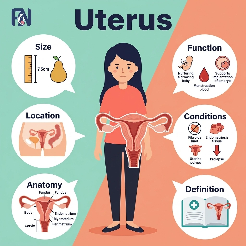
    The uterus refers to the muscular organ in the female pelvis where a fetus develops during pregnancy. In a non-pregnant adult of reproductive age, it measures about 7 to 8 centimeters long and sits in the pelvis between the bladder and rectum, connected to the fallopian tubes, cervix, and vagina. Its anatomy includes distinct regions such as the fundus, body, isthmus, and cervix, along with three tissue layers that support menstruation, implantation, and pregnancy.

    Understanding how the uterus functions and how hormones like estrogen, progesterone, and oxytocin regulate it helps explain everything from a typical menstrual cycle to labor and delivery. Some common uterine conditions are fibroids, adenomyosis, endometriosis, polyps, uterine prolapse, endometrial hyperplasia, and uterine cancer, along with the symptoms that can signal a problem. 

    Monitoring uterine health through regular pelvic exams and timely assessment of symptoms such as heavy bleeding or pelvic pain supports early diagnosis, and treatment options range from medication and hormone therapy to minimally invasive procedures, myomectomy, or hysterectomy depending on the condition.

    What Is the Uterus?

    The uterus is a hollow, muscular reproductive organ located in the female pelvis that houses and nourishes a developing embryo and fetus during gestation. The uterus is commonly called the womb because it serves as the site where a fertilized egg implants and grows throughout pregnancy. 

    Pear-shaped and firm yet flexible in consistency, the uterus is a hormone-responsive organ that changes in size and structure in response to estrogen and progesterone. This responsiveness underlies its central reproductive significance, from monthly menstrual cycles to supporting a full-term pregnancy.

    What Is the Normal Size of the Uterus?

    The normal size of the uterus is about 7–8 cm (2.8–3 inches) long, 4–5 cm (1.6–2 inches) wide, and 2–3 cm (0.8–1.2 inches) thick in a non-pregnant adult of reproductive age. It is typically pear-shaped and weighs approximately 60 grams, with its exact locational position and size varying slightly from person to person. Age, hormonal changes, the phase of the menstrual cycle, and childbirth history can all influence uterine size, with the uterus often appearing slightly larger in women who have given birth.

    Where Is the Uterus Located?

    The uterus is located in the center of the female pelvis, positioned between the bladder in front and the rectum behind. This anatomical position allows the uterus to connect directly with the fallopian tubes at its upper corners and with the cervix and vagina below. 

    The uterus is typically tilted forward, a position known as anteversion, though a backward tilt, or retroversion, is also common and not considered abnormal. Uterine position can vary among individuals without indicating any underlying problem.

    What Is the Anatomy of the Uterus?

    The anatomy of the uterus includes the fundus, body (or corpus), isthmus, and cervix. Each region and layer plays a specific role in supporting menstruation, implantation, and pregnancy. 

    The key anatomical structures of the uterus are listed below.

    • Fundus: The fundus is the rounded, superior portion of the uterus, located above where the fallopian tubes attach on either side. It forms the top of the uterine body and is the widest part of the organ. During pregnancy, doctors measure fundal height from this point to track fetal growth and estimate gestational age.
    • Body (Corpus): The body, or corpus, of the uterus refers to the main, central portion of the organ between the fundus and the isthmus. It is the largest part of the uterus and contains the uterine cavity, the site where implantation occurs. The corpus expands dramatically during pregnancy to accommodate the growing fetus.
    • Isthmus: The isthmus is the narrow region of the uterus located between the body and the cervix. During pregnancy, this area stretches and thins to form the lower uterine segment. Because it thins predictably, the isthmus is also the standard incision site during a cesarean section.
    • Cervix: The cervix refers to the lower, narrow portion of the uterus that connects the uterine cavity to the vagina through the cervical canal. Its inner layer produces cervical mucus, which changes in texture throughout the menstrual cycle to support or block sperm passage. The cervix also plays a critical function during childbirth, dilating fully to allow delivery.

    What Are the Layers of the Uterine Wall?

    The 3 layers of the uterine wall are listed below.

    1. Endometrium: The endometrium is the innermost layer of the uterine wall, made up of a functional layer that sheds monthly and a basal layer that regenerates it. Hormonal changes in estrogen and progesterone drive the functional layer’s growth and shedding during the menstrual cycle. Endometrial glands within this layer support implantation when a fertilized egg arrives.
    2. Myometrium: The myometrium refers to the thick, middle layer of smooth muscle that makes up most of the uterine wall. It enables the uterus to enlarge dramatically during pregnancy and to generate the powerful contractions needed for labor, triggered by oxytocin. After childbirth, the myometrium undergoes postpartum involution, gradually shrinking the uterus back toward its pre-pregnancy size.
    3. Perimetrium: The perimetrium refers to the outer serosal layer that covers the uterus, formed by the visceral peritoneum. It connects the uterus to surrounding pelvic structures and helps anchor its position within the pelvis. This outer layer also contains nerve fibers that contribute to pelvic sensation.

    What Are the Main Components of Uterine Blood Supply, Lymphatics, and Innervation?

    The main components of uterine blood supply, lymphatics, and innervation are listed below.

    • Arterial Supply: The arterial supply of the uterus refers to the network of blood vessels that deliver oxygenated blood to support its function. The uterine artery, a branch of the internal iliac artery, provides the main blood supply, while the ovarian artery contributes additional flow near the fundus. Together, these vessels ensure the uterus receives adequate blood throughout the menstrual cycle and pregnancy.
    • Venous Drainage: Venous drainage of the uterus refers to the system of veins that carry deoxygenated blood away from the organ. The uterine vein serves as the primary vessel for this drainage, running alongside the uterine artery before emptying into the internal iliac vein. Efficient venous drainage helps prevent congestion and supports healthy uterine tissue.
    • Lymphatic Drainage: Lymphatic drainage of the uterus is the network of vessels that carry lymph fluid away from uterine tissue toward the pelvic lymph nodes. This drainage supports immune function and helps clear waste products from the uterine wall. Because uterine cancer can spread through this pathway, pelvic lymph nodes are closely examined during cancer staging.
    • Nerve Supply: The nerve supply of the uterus refers to the network of autonomic nervous system fibers that regulate uterine sensation and muscle activity. These nerves largely originate from the uterovaginal plexus, which carries both sympathetic and parasympathetic input. This nerve supply influences functions such as pain perception and the coordination of contractions during labor.

    What Are the Functions of the Uterus?

    The functions of the uterus include menstruation, implantation, supporting pregnancy, and enabling labor. These functions work together to make conception, pregnancy, and childbirth possible. 

    The main functions of the uterus are listed below.

    • Menstruation: Menstruation is the monthly shedding of the uterine lining, or endometrium, when pregnancy does not occur. The uterus initiates menstruation by shedding its inner lining, the endometrium, when fertilization does not occur. This cyclical process reflects the uterus’s role in hormonal regulation and preparation for potential pregnancy.
    • Implantation: Implantation is the process by which a fertilized egg, now a blastocyst, attaches to the uterine lining about six to ten days after fertilization. Once attached, the blastocyst begins burrowing into the endometrium and initiates the early stages of placental development. Successful implantation marks the true biological start of pregnancy.
    • Pregnancy: The uterus supports pregnancy by expanding dramatically in size to accommodate the growing fetus, from roughly the size of a fist to that of a watermelon. Its muscular walls protect the fetus from outside pressure and impact throughout gestation. Through the placenta, the uterus also facilitates nutrient and oxygen exchange between mother and baby.
    • Labor: The uterus’s role in labor involves powerful myometrial contractions triggered by rising oxytocin levels as childbirth approaches. These contractions gradually cause cervical dilation, widening the cervix to allow the baby to pass through the birth canal. This hormonal and mechanical process culminates in delivery of the baby and, shortly after, the placenta.

    What Are the Hormonal Regulation of the Uterus?

    The hormones that regulate the uterus include estrogen, progesterone, LH, FSH, oxytocin, and prostaglandins.

    The hormones that regulate the uterus are listed below.

    • Estrogen: Estrogen is the hormone that binds to hormone receptors in the uterine lining to drive its growth. It plays a central role in endometrial proliferation during the first half of the menstrual cycle.
    • Progesterone: Progesterone is the hormone released after ovulation that maintains the secretory phase of the endometrium. This phase thickens and stabilizes the uterine lining to support implantation if fertilization occurs.
    • LH: LH, or luteinizing hormone, is the pituitary hormone that surges midcycle to trigger ovulation. It also supports corpus luteum function afterward, which sustains progesterone production for the uterine lining.
    • FSH: FSH, or follicle-stimulating hormone, refers to the pituitary hormone that stimulates follicle development in the ovaries. By promoting follicle growth, FSH indirectly supports the estrogen levels that shape uterine hormone activity.
    • Oxytocin: Oxytocin is the hormone released by the pituitary gland that triggers myometrial contractions during labor. In the uterus, oxytocin released by the pituitary gland stimulates the muscular layer (myometrium) to contract during labor. Its levels rise sharply as delivery approaches, coordinating the rhythmic contractions needed for childbirth.
    • Prostaglandins: Prostaglandins are hormone-like compounds that trigger uterine contractions involved in menstrual shedding and, later in life, cervical ripening before labor. These compounds play a role in uterine function throughout a woman’s lifetime, from her first period through childbirth.

    How Does the Uterus Change Throughout Life?

    The 5 life stages of uterine change are listed below.

    • Childhood: During childhood, the uterus remains small and structurally immature, reflecting typical prepubertal uterus anatomy. Its reproductive function stays inactive until hormonal changes begin later in adolescence.
    • Puberty: During puberty, the uterus undergoes hormonal maturation as rising estrogen levels stimulate its growth. This stage culminates in menarche, the first menstrual period, marking the start of reproductive maturity.
    • Reproductive Years: During the reproductive years, the uterus undergoes monthly cycles of lining growth and shedding tied to ovulation and the menstrual cycle. These cycles prepare the uterus each month to support fertility and potential pregnancy.
    • Pregnancy: During pregnancy, the uterus enlarges dramatically, with increased blood flow supporting placenta formation and ongoing fetal development. It stretches from the size of a fist to fill much of the abdominal cavity by full term.
    • Menopause: During menopause, the uterus responds to hormonal decline and estrogen deficiency with endometrial thinning and gradual uterine atrophy. Maintaining uterine health during this stage still matters, since postmenopausal bleeding can signal an underlying problem.

    How to Maintain Uterine Health?

    The habits that support uterine health are listed below.

    • Schedule Regular Gynecological Exams: Regular gynecological exams, including a pelvic examination, allow doctors to detect uterine conditions early, often before symptoms appear.
    • Get Cervical Cancer Screening: Cervical cancer screening through a Pap smear checks for abnormal cervical cells and is generally recommended every three years for most women.
    • Maintain a Healthy Weight: Maintaining a healthy weight supports balanced hormone levels, since excess body fat can disrupt estrogen levels and increase uterine health risks.
    • Stay Physically Active: Regular physical activity supports hormonal balance and healthy blood flow, both of which benefit reproductive health over time.
    • Practice STI Prevention: STI prevention, including consistent condom use, helps protect against infections that can lead to pelvic inflammatory disease and uterine damage.
    • Quit Smoking: Smoking cessation reduces the risk of several uterine conditions, including cervical cancer and fertility complications linked to reduced blood flow.
    • Attend Prenatal Care Visits: Prenatal care during pregnancy allows doctors to monitor uterine and fetal health, catching complications early for a safer pregnancy.
    • Get the HPV Vaccination: HPV vaccination protects against the human papillomavirus strains most commonly linked to cervical cancer and is recommended before or during early adulthood as part of a healthy lifestyle.

    What Are the Common Uterine Conditions?

    The common uterine conditions include fibroids, adenomyosis, endometriosis, polyps, uterine prolapse, endometrial hyperplasia, and uterine cancer, each affecting the uterus differently and requiring distinct treatment approaches. 

    The common uterine conditions are listed below.

    • Fibroids: Fibroids refer to noncancerous growths that develop within or on the walls of the uterus, varying widely in size and number. They are commonly linked to estrogen sensitivity, genetics, and family history, with risk increasing with age until menopause. Common symptoms include heavy menstrual bleeding, pelvic pressure, and, in some cases, complications with fertility.
    • Adenomyosis: Adenomyosis is a condition in which endometrial tissue grows into the myometrium, the muscular wall of the uterus. Its exact cause is unclear, though risk factors include prior uterine surgery and childbirth. Common symptoms include painful, heavy periods and an enlarged, tender uterus.
    • Endometriosis: Endometriosis is a condition in which tissue similar to the uterine lining grows outside the uterus, often on the ovaries or pelvic tissue. Its causes remain unclear, though genetics and retrograde menstruation are considered contributing risk factors. Common symptoms include pelvic pain and infertility, which can range from mild to severe.
    • Polyps: Uterine polyps refer to small, usually noncancerous growths that develop from the endometrial lining and extend into the uterine cavity. They are linked to excess estrogen exposure and become more common with age, particularly around menopause. The most common symptom is abnormal bleeding, including spotting between periods.
    • Uterine Prolapse: Uterine prolapse refers to a condition in which the uterus slips down into or out of the vaginal canal due to weakened support structures. It is commonly caused by pelvic floor weakness following childbirth, aging, or chronic straining. Symptoms often include a sensation of pelvic heaviness or a visible bulge at the vaginal opening.
    • Endometrial Hyperplasia: Endometrial hyperplasia is abnormal thickening of the uterine lining, usually caused by excess estrogen without enough progesterone to balance it. Risk factors include obesity, polycystic ovary syndrome, and irregular ovulation. The most common symptom is abnormal uterine bleeding, which requires evaluation since it can sometimes precede cancer.
    • Uterine Cancer: Uterine cancer refers to a malignant growth that typically begins in the endometrial lining of the uterus. Risk factors include obesity, prolonged estrogen exposure, and a family history of the disease. Common symptoms include abnormal vaginal bleeding, particularly after menopause, along with pelvic pain in more advanced cases.

    What Symptoms Can Indicate a Uterine Problem?

    The common symptoms that may indicate a uterine problem are listed below.

    • Heavy Menstrual Bleeding: Heavy menstrual bleeding refers to periods with unusually heavy flow or prolonged duration. It can be a sign of fibroids, polyps, or hormonal imbalance.
    • Pelvic Pain: Pelvic pain refers to discomfort in the lower abdomen that can range from mild to severe. It can result from fibroids, adenomyosis, or endometriosis.
    • Infertility: Infertility refers to the inability to conceive after a year of regular, unprotected intercourse. Structural or hormonal uterine problems can prevent conception by disrupting implantation.
    • Abnormal Bleeding: Abnormal bleeding refers to vaginal bleeding that occurs outside a normal menstrual pattern. It includes bleeding between periods or after intercourse and may signal an underlying condition.
    • Postmenopausal Bleeding: Postmenopausal bleeding refers to any vaginal bleeding that occurs after menopause has been confirmed. It always requires prompt medical evaluation to rule out uterine cancer.
    • Pelvic Pressure: Pelvic pressure means a heavy or full sensation in the lower abdomen or pelvis. It is commonly associated with fibroids or uterine prolapse.
    • Painful Intercourse: Painful intercourse refers to discomfort or sharp pain felt during or after sexual activity. It can result from endometriosis, adenomyosis, or pelvic inflammation.
    • Abnormal Discharge: Abnormal discharge means vaginal discharge that differs in color, odor, or consistency from what is typical. It may signal infection, polyps, or, rarely, uterine cancer.
    • Recurrent Miscarriage: Recurrent miscarriage refers to the loss of two or more consecutive pregnancies before 20 weeks. Uterine abnormalities, such as a septum or fibroids, can be an underlying cause.

    How Are Uterine Conditions Diagnosed?

    The common methods used to diagnose uterine conditions are listed below.

    • Pelvic Examination: A pelvic examination is a physical exam in which a doctor checks the uterus, cervix, and surrounding structures. It includes a bimanual examination and a speculum examination to assess size, shape, and tenderness.
    • Ultrasound: An ultrasound, including transabdominal and transvaginal ultrasound, is an imaging test that uses sound waves to create images of the uterus. It helps visualize the uterus and detect abnormalities such as fibroids, polyps, or thickened lining.
    • MRI: A pelvic MRI is an imaging test that uses magnetic fields to create highly detailed pictures of pelvic structures. It provides detailed soft tissue imaging of the uterus, helping distinguish between different types of masses.
    • CT Scan: A CT scan, or computed tomography, is an imaging test that combines multiple X-ray images to create cross-sectional pictures of the pelvis. It provides pelvic imaging often used to assess whether uterine cancer has spread.
    • Hysteroscopy: Hysteroscopy is a procedure in which a thin, lighted scope is inserted through the cervix into the uterus. It allows direct visualization of the uterine cavity to detect and sometimes treat polyps or fibroids.
    • Endometrial Biopsy: An endometrial biopsy is a procedure that involves taking a small tissue sample from the uterine lining. This sample undergoes histopathology to detect abnormal cells and helps guide decisions about appropriate treatments.

    What Are the Best Treatments for Uterine Conditions in India?

    The best treatments for uterine conditions in India include medications, hormone therapy, minimally invasive procedures, myomectomy, and hysterectomy, available at major hospitals across the country. 

    The best treatments for uterine conditions in India are listed below.

    • Delhi: Delhi offers advanced gynecological centers providing medications, hormone therapy, and minimally invasive procedures such as laparoscopic myomectomy, with treatment cost-range varying by hospital and procedure.
    • Mumbai: Mumbai is home to leading hospitals performing myomectomy and hysterectomy alongside fertility-focused uterine care, with a wide cost range depending on complexity.
    • Bangalore: Bangalore provides access to gynecologists specializing in minimally invasive procedures and hormone therapy for conditions such as fibroids and endometriosis.
    • Chennai: Chennai has established centers offering both medical management and surgical treatments, including myomectomy and hysterectomy, for a range of uterine conditions.
    • Hyderabad: Hyderabad offers well-equipped hospitals providing hormone therapy and minimally invasive procedures, with treatment costs varying based on diagnosis and approach.
    • Kolkata: Kolkata has growing gynecological infrastructure with specialists treating fibroids, adenomyosis, and other uterine conditions through medication or surgery.
    • Pune: Pune provides access to gynecological specialists and diagnostic centers offering treatment options ranging from medication to hysterectomy, depending on condition severity.

    How Much Does Uterine Condition Treatment Cost in India?

    The cost of uterine condition treatment in India ranges from ₹15,000 to ₹4,00,000 depending on the diagnosis, treatment approach, and hospital chosen. Initial consultations and diagnostic tests generally cost between ₹500 and ₹2,500 per visit. Treatment costs vary further by approach, with medication and hormone therapy at the lower end and minimally invasive procedures, myomectomy, or hysterectomy at the higher end.

    Can Uterine Conditions Cause Infertility?

    Yes, uterine conditions can cause infertility because they can disrupt implantation, hormone balance, or the shape of the uterine cavity needed for conception. These conditions interfere with the uterus’s ability to support a fertilized egg or maintain a healthy pregnancy. Common underlying conditions associated with infertility include fibroids, adenomyosis, endometriosis, and polyps, all of which can directly affect reproductive health.

    How to Find the Right Treatments in India for Infertility Caused by Uterine Conditions?

    To find the right infertility treatment in India for uterine conditions, compare experienced fertility specialists, recommended treatments, and treatment costs based on your diagnosis and reproductive needs.

    The right treatment depends on the anatomy of the uterus, its functions, hormonal regulation, the size and location of the uterus, and the specific uterine conditions affecting fertility. Important treatments include medications, hysteroscopic surgery, myomectomy, treatment for uterine polyps or adhesions, and fertility treatments such as IUI or IVF when recommended. 

    Before choosing a clinic, check the accuracy of the diagnosis, specialist expertise, success rates, available facilities, follow-up care, and treatment cost. Fertility Network helps you find affordable fertility treatment options near you in India by connecting you with trusted fertility specialists and clinics.

  • Fallopian Tubes (Uterine Tubes): Definition, Anatomy, Function, Conditions and Treatments

    Fallopian Tubes (Uterine Tubes): Definition, Anatomy, Function, Conditions and Treatments

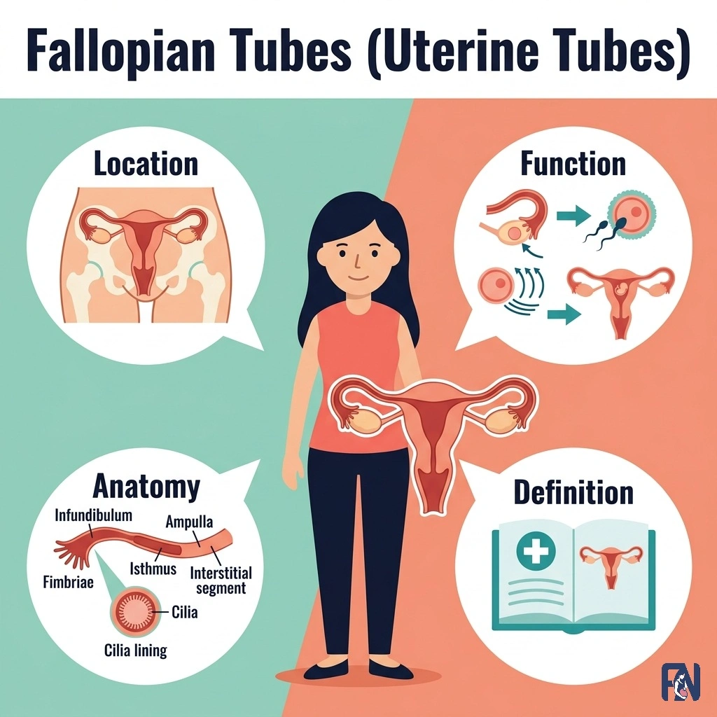
    The fallopian tubes refer to the paired ducts that carry eggs from the ovaries to the uterus. They are essential for reproduction because fertilization normally occurs within them, and they also serve as the pathway through which a fertilized egg travels to the uterus for implantation. Understanding fallopian tube anatomy and function helps explain how natural conception happens and why blocked or damaged tubes are a leading cause of infertility.

    Some common fallopian tube conditions include blocked tubes, hydrosalpinx, pelvic inflammatory disease, endometriosis, ectopic pregnancy, and tubal cancer. Monitoring tubal health and seeking prompt medical evaluation for symptoms such as pelvic pain or irregular bleeding supports early diagnosis and treatment. Treatment options range from medication and minimally invasive procedures to tuboplasty, salpingostomy, salpingectomy, or IVF, depending on the condition and its severity.

    What Are the Fallopian Tubes?

    The fallopian tubes, also known as uterine tubes or oviducts, are paired, bilateral muscular ducts that extend from the uterus toward each ovary, providing the pathway through which the oocyte, sperm, and early embryo travel. Fallopian tubes measure 10 to 14 centimeters in length, with a diameter ranging from 1 to 10 millimeters depending on the segment. 

    Anatomically, these hollow muscular tubes are located in the upper female pelvis, where they extend laterally from the uterus toward the ovaries as part of the female reproductive tract. Although the fallopian tubes are closely associated with the ovaries, they are not directly attached to them. Instead, the open funnel-shaped end of each tube lies near the corresponding ovary to capture the oocyte after ovulation.

    The fallopian tubes provide the pathway for the oocyte to travel from the ovary toward the uterus. They also serve as the usual site of fertilization, where sperm and the oocyte typically meet before the developing embryo is transported to the uterus for implantation.

    What Is the Anatomy of the Fallopian Tubes?

    The anatomy of the fallopian tubes includes the peritoneal covering, the broad ligament, and the mesosalpinx that support and suspend each tube within the pelvis. 

    The anatomical structures of the fallopian tube are listed below.

    • Peritoneum: The peritoneum is the thin membrane that covers the outer surface of each fallopian tube, forming part of its serosal layer. It provides a smooth surface that reduces friction against surrounding pelvic organs. This covering also connects the tube to the broader peritoneal cavity.
    • Broad Ligament: The broad ligament is a wide fold of peritoneum that supports the uterus, fallopian tubes, and ovaries within the pelvis. It anchors the fallopian tube along its length and helps maintain its position relative to the uterus. The ligament also carries blood vessels and nerves toward the reproductive organs.
    • Mesosalpinx: The mesosalpinx is the upper portion of the broad ligament that specifically suspends and supports the fallopian tube. It carries the blood vessels, including branches of the ovarian and uterine arteries, that supply the tube. The mesosalpinx also provides a degree of mobility that helps the tube capture the egg during ovulation.

    How Does the Fallopian Tube Blood Supply, Nerve Supply, and Lymphatic Drainage Work?

    The fallopian tube blood supply, nerve supply, and lymphatic drainage work together to deliver oxygen and nutrients, regulate tubal function, and remove excess tissue fluid from these important parts of the reproductive tract. Blood supply comes from a dual arterial source, the ovarian and uterine arteries, which form a protective anastomosis within the mesosalpinx, while venous drainage follows a parallel path. 

    Nerve supply comes from autonomic innervation via the ovarian and inferior hypogastric plexuses, with sympathetic and parasympathetic fibers regulating tubal motility and pain sensation. Lymphatic drainage flows primarily to the lumbar, or paraaortic, lymph nodes, with some drainage from the medial portion reaching the internal iliac lymph nodes.

    What Are the Important Parts of the Fallopian Tube?

    The important parts of the fallopian tube are listed below.

    • Fimbriae: The fimbriae refer to the finger-like projections at the tube’s outer end near the ovary. These projections sweep over the ovary’s surface to capture the ovulated egg during ovulation. Their clinical significance lies in their vulnerability to damage from infection or endometriosis, which can impair egg capture.
    • Infundibulum: The infundibulum refers to the funnel-shaped opening of the fallopian tube nearest the ovary. It transitions directly from the fimbriae and serves as the entry point for the egg after capture. This funnel shape helps guide the egg smoothly into the rest of the tube.
    • Ampulla: The ampulla is the longest and widest section of the fallopian tube, located between the infundibulum and isthmus. It is the typical site of fertilization, where sperm meets egg, and early embryo development begins. Its wide lumen and rich ciliary lining support this critical reproductive process.
    • Isthmus: The isthmus of the fallopian tube refers to the narrow, muscular segment closest to the uterus. This segment transports the egg or early embryo toward the uterus through peristalsis, a wave-like muscular contraction. Its narrow diameter also makes it a common site for tubal blockage.
    • Intramural (Interstitial) Segment: The intramural, or interstitial, segment refers to the portion of the fallopian tube that passes through the muscular wall of the uterus. It serves as the entry point into the uterine cavity for the egg or embryo. Its clinical importance includes relevance to tubal ligation procedures and interstitial ectopic pregnancies, which can affect histology and surgical planning.

    What Is the Histology of the Fallopian Tubes?

    The histology of the fallopian tubes refers to the three distinct layers, including the mucosa, muscularis, and serosa, that give the tube its structure and functions. The mucosa lines the inner lumen, the muscularis provides the smooth muscle needed for peristaltic movement, and the outer serosa offers a protective covering. 

    Within the mucosa, ciliated cells beat rhythmically to help move the ovum along the tube, while secretory peg cells release fluid that nourishes the ovum and supports sperm function. This histological organization is significant because it directly supports ovum transport, fertilization, and early reproductive function.

    What Are the Functions of the Fallopian Tubes?

    The functions of the fallopian tubes include capturing the egg, transporting it toward the uterus, supporting fertilization, and moving the developing embryo onward for implantation. 

    The main functions of the fallopian tubes are listed below.

    • Egg Capture: Egg capture refers to the process by which the fimbriae sweep over the ovary to catch the oocyte released during ovulation. Coordinated muscular movement draws the fimbriae close to the ovarian surface at the moment of egg release. This capture step is essential, since a released oocyte cannot travel to the uterus on its own.
    • Egg Transport: Egg transport is the movement of the egg along the fallopian tube toward the uterus after capture. Rhythmic beating of cilia lining the tube, combined with peristalsis from smooth muscle contractions, drives this tubal motility. Together, these mechanisms ensure the egg reaches the ampulla in time for fertilization.
    • Fertilization: Fertilization refers to the union of sperm and egg, which typically occurs within the ampulla of the fallopian tube. Sperm undergo capacitation and the acrosome reaction during their migration through the reproductive tract, allowing one sperm to penetrate the egg. This union forms a zygote, marking the earliest stage of a successful pregnancy and the foundation of fertility.
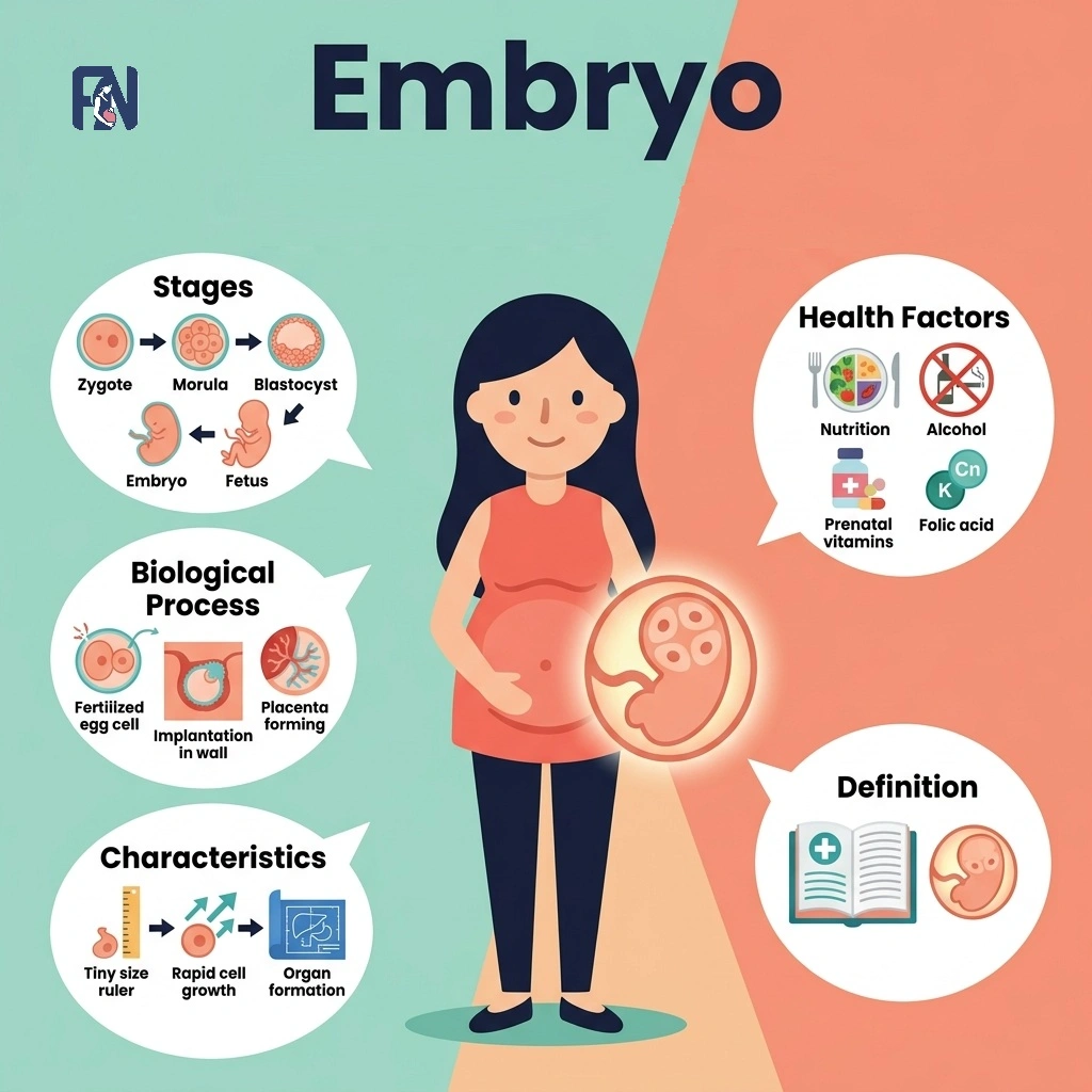
    • Embryo Transport: Embryo transport is the movement of the developing embryo through the fallopian tube toward the uterus following fertilization. As the embryo travels, it undergoes cleavage, dividing repeatedly until it reaches the blastocyst stage. The embryo typically arrives in the uterus about three to five days after fertilization, implanting into the endometrium shortly after, a process essential to fertility.

    What Is the Role of Uterine Tubes in Fertility?

    The fallopian tubes play a role in fertility by providing the pathway where fertilization occurs and by transporting the egg and embryo toward the uterus. Natural conception depends on patent, or open, tubes, since blockage or damage can prevent sperm and egg from meeting, a condition known as tubal factor infertility. Healthy cilia are equally important, since impaired ciliary function from various uterine tube conditions can slow or halt egg and sperm transport even when the tubes remain open.

    What Are the Common Fallopian Tube Conditions?

    The common fallopian tube conditions include blocked fallopian tubes, hydrosalpinx, pelvic inflammatory disease, endometriosis, ectopic pregnancy, and tubal cancer, each affecting the tubes in different ways. 

    The common disorders of the fallopian tubes are listed below.

    • Blocked Fallopian Tubes: Blocked fallopian tubes refer to a tubal obstruction that prevents the egg and sperm from meeting, a leading cause of infertility. Common causes include pelvic infections, endometriosis, and adhesions from prior surgery, all of which can scar or narrow the tube. This tubal blockage is often asymptomatic and typically discovered only through imaging tests such as an HSG during a fertility evaluation.
    • Hydrosalpinx: Hydrosalpinx refers to a condition in which a fallopian tube becomes blocked and fills with fluid accumulation, usually following infection or prior pelvic surgery. This fluid can leak into the uterus and interfere with embryo implantation, reducing natural fertility. Because of this impact, doctors often recommend salpingectomy, or tube removal, before pursuing IVF.
    • Pelvic Inflammatory Disease: Pelvic inflammatory disease, or PID, refers to an infection of the reproductive organs most often caused by untreated chlamydia or gonorrhea. Left untreated, PID can cause tubal scarring and adhesions that damage the fallopian tubes permanently. Prevention strategies include safe sex practices, regular STI screening, and prompt treatment of any diagnosed infection.
    • Endometriosis: Endometriosis refers to a condition in which tissue similar to the uterine lining grows outside the uterus, sometimes affecting the fallopian tubes directly. This growth can create scar tissue and pelvic adhesions that distort normal tubal anatomy. These changes can affect fertility by blocking the tube or impairing its ability to capture and transport the egg.
    • Ectopic Pregnancy: An ectopic pregnancy is a tubal pregnancy in which a fertilized egg implants inside the fallopian tube instead of the uterus. Risk factors include prior tubal damage, PID, and previous ectopic pregnancy, and symptoms often include sharp abdominal pain and abnormal bleeding. Because rupture can cause life-threatening internal bleeding, this condition requires emergency treatment, often with methotrexate or surgery.
    • Tubal Cancer: Tubal cancer refers to primary fallopian tube carcinoma, a rare gynecologic cancer that originates within the tube itself. It is more common in women with a BRCA mutation, though it can occur without a known genetic cause. Symptoms are often vague, including pelvic pain or abnormal discharge, and diagnosis typically requires imaging alongside surgical evaluation.

    What Are the Symptoms of Fallopian Tube Problems?

    The symptoms of fallopian tube problems are listed below.

    • Pelvic Pain: Pelvic pain is discomfort in the lower abdomen that can signal infection, endometriosis, or a blocked tube.
    • Infertility: Infertility is the inability to conceive, often the first noticeable sign of tubal blockage or damage that prevents healthy egg transport or fertilization.
    • Painful Intercourse: Painful intercourse is discomfort during sex that can result from adhesions or inflammation affecting the tubes.
    • Irregular Bleeding: Irregular bleeding is unpredictable spotting or bleeding that may accompany infection or an ectopic pregnancy.
    • Chronic Pelvic Pain: Chronic pelvic pain is persistent discomfort lasting months, often linked to scarring or endometriosis affecting the tubes.
    • Vaginal Discharge: Vaginal discharge that is unusual in color or odor can indicate an underlying infection such as PID.
    • Fever: Fever is a common sign of active infection, such as pelvic inflammatory disease affecting the tubes.
    • Ectopic Pregnancy Symptoms: Ectopic pregnancy symptoms include sharp abdominal pain, shoulder pain, and dizziness, requiring immediate emergency care.

    How to Keep Fallopian Tubes Healthy?

    To keep uterine tubes healthy, follow the tips listed below.

    • Practice Safe Sex: Practicing safe sex reduces the risk of sexually transmitted infections that can lead to PID and tubal damage.
    • Get Regular STI Screening: Regular STI screening supports prevention by diagnosing infections early, before they can cause lasting tubal scarring.
    • Seek Prompt Treatment for Infections: Seeking prompt treatment for pelvic infections helps prevent them from progressing to PID or tubal scarring.
    • Manage Endometriosis Early: Managing endometriosis early with medical care can help limit adhesions that might otherwise distort tubal anatomy.
    • Schedule Regular Gynecological Exams: Regular gynecological exams allow early detection of tubal or pelvic conditions before symptoms become severe.

    How to Diagnose Fallopian Tube Disorders?

    Doctors diagnose fallopian tube conditions using imaging tests, minimally invasive procedures, and direct visualization methods that assess tubal shape and patency. 

    The common methods used to diagnose fallopian tube conditions are listed below.

    • Hysterosalpingography (HSG): Hysterosalpingography, or HSG, is an X-ray procedure that uses contrast dye to check whether the fallopian tubes are open. It is commonly used during a fertility workup to detect tubal blockage or abnormalities in the uterine cavity. Doctors typically recommend HSG early in an infertility evaluation, usually shortly after menstruation ends.
    • HyCoSy: HyCoSy is an ultrasound-based procedure that uses a saline or foam contrast solution to evaluate tubal patency. It offers a radiation-free alternative to HSG and can be performed in a standard gynecology clinic. Doctors often recommend HyCoSy for women who prefer to avoid X-ray exposure during a fertility evaluation.
    • Ultrasound: An ultrasound is an imaging test that uses sound waves to visualize the uterus, ovaries, and surrounding pelvic structures. It helps identify conditions such as hydrosalpinx or ovarian cysts, though it cannot directly confirm tubal patency. Doctors typically use ultrasound as an initial step before more specialized tubal testing.
    • MRI: A pelvic MRI is an imaging test that uses magnetic fields to produce detailed pictures of the fallopian tubes and surrounding tissue. It is particularly useful for evaluating complex cases, such as suspected tubal cancer or deep endometriosis. Doctors typically recommend MRI when ultrasound findings are unclear or need further clarification.
    • Diagnostic Laparoscopy: Diagnostic laparoscopy is a minimally invasive surgical procedure that allows direct visualization of the fallopian tubes and pelvic organs. It can confirm blockage, adhesions, or endometriosis while also allowing early treatment during the same procedure. Doctors typically recommend diagnostic laparoscopy when other imaging tests are inconclusive or when surgical correction may be needed.

    What Are the Best Treatments for Fallopian Tube Conditions in India?

    The best treatments for fallopian tube conditions in India include medications, tubal recanalization, tuboplasty, salpingostomy, salpingectomy, and in vitro fertilization, available at major fertility centers across the country. 

    The best treatments for fallopian tube conditions in India are listed below.

    • Medications: Medications, such as antibiotics, are used to treat infections like PID before they progress to permanent tubal scarring. This treatment sits at the lower end of the cost range compared to surgical options. It works best when started early, before significant tubal damage occurs.
    • Tubal Recanalization: Tubal recanalization is a minimally invasive procedure that reopens a blocked segment of the fallopian tube using a catheter. It is typically used for blockages near the uterine end of the tube. This procedure can restore natural fertility in appropriate candidates without more invasive surgery.
    • Tuboplasty: Tuboplasty is a surgical procedure that repairs or reconstructs a damaged fallopian tube to restore its function. It is often used for blockages caused by adhesions or scar tissue elsewhere along the tube. Success depends on the extent and location of the damage.
    • Salpingostomy: Salpingostomy is a surgical procedure that creates a new opening in a blocked fallopian tube, often at the fimbrial end. It is commonly used to treat hydrosalpinx or distal tubal blockage. This procedure aims to restore tubal function, though success rates vary with the severity of damage.
    • Salpingectomy: Salpingectomy is the surgical removal of a damaged or diseased fallopian tube, often performed for severe hydrosalpinx or ectopic pregnancy. It is frequently recommended before IVF when a damaged tube is likely to reduce implantation success. Removing the affected tube does not eliminate fertility, since the remaining tube or IVF can still support conception.
    • In Vitro Fertilization (IVF): In vitro fertilization, or IVF, bypasses the fallopian tubes entirely by fertilizing eggs outside the body and transferring the embryo directly into the uterus. It is often recommended when tubal damage is severe or when other treatments have not restored fertility. IVF costs fall at the higher end of the cost range compared to medication or minor procedures.

    How Much Does Fallopian Tube Condition Treatment Cost in India?

    The cost of fallopian tube condition treatment in India ranges from ₹25,000 to ₹1,60,000 depending on the diagnosis, treatment approach, and hospital chosen. Consultations and diagnostic tests, including HSG or ultrasound, typically add a few thousand rupees to the overall cost. Treatment costs vary further by approach, with medication at the lower end and minimally invasive procedures, surgery, or IVF for fertility restoration at the higher end.

    Can Fallopian Tube Conditions Cause Infertility?

    Yes, fallopian tube conditions can cause infertility because they can block or damage the tubes needed for natural conception. When the tubes cannot allow sperm and egg to meet or cannot transport a fertilized egg to the uterus, pregnancy becomes difficult or impossible without intervention. Common underlying conditions associated with fallopian tube problems include blocked tubes, hydrosalpinx, pelvic inflammatory disease, and endometriosis, all of which can directly affect reproductive health and contribute to infertility.

    How to Get Proper Treatments in India for Infertility Caused by Fallopian Tube Conditions?

    To get proper treatments in India for infertility caused by fallopian tube conditions, consult a qualified fertility specialist for an accurate diagnosis of the uterine tube anatomy and functions before choosing the most appropriate treatment.

    The recommended treatment depends on the underlying condition and may include medications for infections, laparoscopic surgery to repair selected tubal abnormalities, or assisted reproductive techniques such as in vitro fertilization (IVF) when tubal damage is severe. Before starting treatment, check the exact diagnosis, the extent of tubal damage, the condition of the other reproductive organs, the specialist’s experience, success rates, available treatment options, and the total treatment cost.

    At Fertility Network, we help you compare affordable fertility treatment options and connect you with trusted fertility specialists and clinics near you across India, making it easier to find quality care that fits your medical needs and budget.

  • Embryo: Definition, Development Stages, Characteristics and Health Factors

    Embryo: Definition, Development Stages, Characteristics and Health Factors

    An embryo refers to an early-stage multicellular organism developing from a fertilized egg before major body systems form. The main stages of human embryonic development include zygote, blastocyst, embryo, and fetus, marking critical transitions in prenatal growth. Embryo formation begins biologically with fertilization, the union of sperm and egg, and proceeds through rapid cell division, implantation, and the progressive differentiation of tissues. Important characteristics of an embryo include cellular totipotency, rapid mitotic division, bilateral symmetry, and the early formation of organ rudiments. 

    The embryo developmental process includes blastocyst formation by day 5, uterine implantation by the end of week 2, and the completion of major organ structures by week 8. The main factors affecting embryo health include maternal age, genetic composition, hormonal balance, nutritional status, and environmental exposures, each of which can contribute to developmental abnormalities when disrupted. 

    Understanding embryonic health is fundamental to preventing pregnancy complications and improving reproductive outcomes for families affected by infertility. Fertility Network connects patients across India with qualified fertility specialists and evidence-based treatments to support healthy embryonic development at every stage of the process.

    What Is an Embryo?

    An embryo is a multicellular prenatal organism at its earliest stage of development organism, formed immediately after fertilization of an egg by sperm and developing continuously through the eighth week of gestation.

    The difference between an animal embryo and a human embryo is that animal embryos may develop externally within eggs or inside various anatomical structures, whereas a human embryo develops exclusively within the uterus following implantation into the endometrial lining. The embryo forms when fertilization produces a single-celled zygote that undergoes successive mitotic divisions, generating a progressively more complex multicellular structure over the following days and weeks. 

    The term “embryo” in its modern medical usage was formally established in the 19th century as anatomical researchers began systematically distinguishing prenatal developmental stages and describing the structural sequence from fertilization onward. According to the presentation data from the American Society for Reproductive Medicine (ASRM) showed that incubating eggs in specialized, nutrient-dense media for just 3 to 6 hours before fertilization significantly improved overall embryo development, independent of maternal age.

    What is The Location of an Embryo?

    The location of an embryo is inside the uterus (womb), where it attaches to the endometrial lining and continues to develop across the different stages of early intrauterine pregnancy. The fertilized egg attaches to the uterine lining, known as the endometrium, after migrating from the fallopian tube, and this attachment marks the true beginning of the embryonic stage proper. 

    Implantation commonly occurs in the upper portion of the uterus, called the fundus, where a richly vascularized environment supplies the hormonal signals and nutrients that sustain the embryo through the critical early weeks of development.

    What is the Duration of the Full Embryonic Development?

    The duration of the full embryonic development is from fertilization through the 8 weeks of pregnancy, or approximately 56 days, during which all primary organ systems are established within the developing organism inside the uterine environment. 

    The embryonic period begins with the process of fertilization and continues until the completion of the eighth week of development, marked by rapid cell division, tissue formation, and the development of major organs and body structures that define the human body plan. Following the embryonic stage, development transitions into the fetal stage, during which all established organ systems grow, mature, and prepare for postnatal independent function outside the womb.

    What are the Stages of Human Embryonic Development?

    The stages of human embryonic development include fertilization, cleavage, blastocyst formation, implantation, gastrulation, organogenesis, and the fetal stage, progressing from a single-cell zygote to a fully developed baby.

    The stages of human embryonic development are listed below.

    1. Fertilization Stage (12–24 Hours Post-Ovulation): The fertilization stage refers to the biological union of a sperm cell and an oocyte in the ampullary region of the fallopian tube, producing a single diploid zygote that carries the complete and unique chromosomal arrangement of both parents. This newly formed zygote contains all genetic information required to direct every subsequent stage of embryonic development. Fertilization marks the biological origin point of a new human organism and initiates the cascade of cellular events that follow.
    2. Cleavage Stage (Days 1–4): The cleavage stage refers to a rapid series of mitotic divisions of the zygote that produce progressively smaller daughter cells called blastomeres, without any net increase in the total cellular volume of the developing mass. This process begins approximately 24 hours after fertilization and continues through day 4, transforming the single-celled zygote into a compact, mulberry-shaped multicellular structure known as the morula. The cleavage stage establishes the cellular foundation upon which all subsequent structural differentiation depends.
    3. Blastocyst Stage (Days 5–7): The blastocyst stage refers to the phase in which the morula undergoes blastulation, transforming into a hollow, fluid-filled structure characterized by an internal cavity called the blastocoel, an outer trophoblast layer, and an inner cell mass of pluripotent cells. The trophoblast layer initiates the biochemical preparation for uterine implantation, while the inner cell mass contains all cells destined to form the embryo itself. This stage concludes the pre-implantation period and positions the developing structure for physical attachment to the maternal endometrium.
    4. Implantation Stage (Days 6–7 to End of Week 2): The implantation stage refers to the process by which the blastocyst embeds itself into the endometrial lining of the uterus, initiating a physical and biochemical connection between the embryo and the maternal body. This process begins around days 6 to 7 post-fertilization and is typically completed by the end of the second week of development. Successful implantation establishes the nutritional and hormonal exchange essential for all sustained embryonic development that follows.
    5. Gastrulation Stage (Weeks 2–3): The gastrulation stage refers to the reorganization of the bilaminar embryonic disc into three primary germ layers, ectoderm, mesoderm, and endoderm, occurring between weeks 2 and 3 of development. Each germ layer serves as the developmental source of distinct organ systems, collectively establishing the foundational body plan of the entire organism. Gastrulation transforms a structurally simple disc into a trilaminar embryo with defined axes, polarity, and tissue identity.
    6. Organogenesis Stage (Weeks 3–8): The organogenesis stage refers to the critical developmental period during which the three primary germ layers differentiate into organ rudiments and specialized tissues, driving the formation of all major body systems including the neural tube, cardiac structures, limb buds, and the digestive, respiratory, and urogenital systems. This stage spans weeks 3 through 8 of the embryonic period and represents the most teratogenically sensitive window of human development, as all primary organ systems are actively forming. Organogenesis concludes with the structural completion of all major organs and marks the biological transition from embryo to fetus.
    7. Fetal Stage (Week 9 to Birth): The fetal stage refers to the developmental period beginning at week 9 and continuing through delivery, defined by progressive organ maturation, functional refinement, and overall growth rather than the primary formation of new structures. All organ systems established during organogenesis undergo continuous specialization and physiological preparation for extrauterine life throughout this period. The fetal stage represents the longest phase of prenatal development and culminates in a fully formed organism capable of independent survival at birth.

    What Is the Difference Between Embryo and Fetus?

    The difference between an embryo and a fetus is that an embryo is the early stage, up to 8 weeks post-fertilization, while a fetus is the later stage from the 9th week until birth.

    An embryo becomes a fetus at the conclusion of the eighth gestational week, once the foundational architectural organization of the human body has been structurally established and all primary organs have formed. The characteristics of an embryo transition into a fetus as organ formation gives way to organ growth and maturation, with the fetal period marked by increasing body size, the refinement of organ systems, and physiological preparation for independent life outside the womb.

    What are The Characteristics of an Embryo?

    The characteristics of an embryo include rapid mitotic division, cellular totipotency and pluripotency, bilateral symmetry, organ rudiment formation, developmental plasticity, and dependence on the uterine environment.

    The characteristics of an embryo are listed below.

    • Rapid Mitotic Division: Rapid mitotic division refers to the accelerated rate of cellular multiplication in which the zygote progresses from a single cell to a complex multicellular organism within days following fertilization. This process generates the necessary cellular mass for tissue specification and subsequent organ formation throughout the embryonic stage.
    • Cellular Totipotency and Pluripotency: Cellular totipotency and pluripotency refer to the extraordinary capacity of early embryonic cells to differentiate into any cell type present in the human body, a defining biological characteristic of the earliest stages of embryonic development. Totipotency is characteristic of the initial cleavage-stage cells, while pluripotency becomes the dominant property within the inner cell mass of the blastocyst.
    • Bilateral Symmetry: Bilateral symmetry refers to the structural organization of the developing embryo in which a definable left-right axis emerges along the central body plane during gastrulation, establishing the architectural template for the placement of all major organ systems. This feature governs the organized and reproducible arrangement of developing organs within the growing embryonic body.
    • Organ Rudiment Formation: Organ rudiment formation refers to the emergence of early structural precursors to the major organ systems, including the neural tube, cardiac loops, and limb buds, during the period from weeks 3 through 8 of embryonic development. These rudiments serve as the biological foundation from which fully differentiated and functionally mature organs will subsequently develop during the fetal period.
    • Developmental Plasticity: Developmental plasticity refers to the embryo’s capacity to adjust its developmental trajectory in response to environmental signals, epigenetic regulatory mechanisms, or disruptions in maternal physiology encountered during critical windows of organogenesis. Developmental plasticity affects the embryo by modifying its growth in response to environmental signals. While this flexibility enables adaptive responses, it also creates vulnerability, as external perturbations during sensitive developmental periods can permanently alter normal embryonic growth patterns.
    • Dependence on Uterine Environment: Dependence on the uterine environment refers to the embryo’s complete reliance on the endometrium and its associated vascular supply for oxygen, nutrients, and the hormonal signals required to sustain its development during the earliest weeks of pregnancy. Inadequate uterine receptivity, compromised endometrial vascularization, or hormonal insufficiency can impair the embryo’s capacity to implant and develop normally.

    What Factors Affect Embryo Health?

    The factors that affect an embryo include maternal age, genetic composition, hormonal balance, nutritional status, environmental exposures, and uterine health.

    The factors that affect an embryo are listed below.

    • Maternal Age: Maternal age refers to the chronological age of the mother at conception, which directly influences the chromosomal integrity of the oocyte and the developmental quality of the embryo produced following fertilization. Advanced maternal age is associated with significantly increased rates of chromosomal abnormalities and reduced embryo viability and implantation success.
    • Genetic Composition: Genetic composition refers to the complete inherited genetic material contributed by both parents, which determines the embryo’s developmental blueprint and its susceptibility to inherited disorders or abnormalities in cellular function and tissue organization. Mutations, chromosomal rearrangements, or gene deletions in parental genetic material can be transmitted directly to the embryo and disrupt normal developmental signaling pathways.
    • Hormonal Balance: Hormonal balance refers to the precise regulation of reproductive hormones, including estrogen, progesterone, and human chorionic gonadotropin, that collectively orchestrate endometrial preparation, embryo implantation, and the early physiological support of the developing embryo. Disruptions in these hormonal signals can compromise endometrial receptivity and lead to embryo developmental abnormalities, implantation failure, or early pregnancy loss.
    • Nutritional Status: Nutritional status refers to the adequacy of the mother’s intake of essential micronutrients, including folate, iron, iodine, and omega-3 fatty acids, that are required to support the biochemical processes underlying embryonic cell division, organogenesis, and tissue development. A clinical study published in Reproductive BioMedicine Online (2026) found that women who adhered to a strict pro-fertility diet combined with daily omega-3 fatty acid supplementation for 60 days significantly increased their fertilization rates. It also led to a higher proportion of top-tier Grade A and B embryos. Nutritional deficiencies during this period are linked to neural tube defects, intrauterine growth restriction, and other developmental abnormalities.
    • Environmental Exposures: Environmental exposures refer to teratogenic agents, including alcohol, tobacco, certain medications, ionizing radiation, and industrial chemicals, that can interfere with normal embryonic cellular division and differentiation when encountered during sensitive developmental windows. Even brief or limited exposure to specific teratogens during the embryonic period can produce lasting structural or functional abnormalities in the developing organ systems.
    • Uterine Health: Uterine health refers to the structural and functional integrity of the uterus, encompassing the quality of the endometrial lining and the absence of anomalies such as fibroids, polyps, or intrauterine adhesions that may obstruct implantation or impair embryo development. A compromised uterine environment reduces the embryo’s capacity to implant successfully and receive the sustained nourishment required for healthy continued development.

    What are The Common Disorders and Developmental Abnormalities of an Embryo?

    Common disorders and developmental abnormalities include genetic abnormalities, chromosomal conditions, and developmental defects, each of which can compromise the integrity and trajectory of embryonic development in clinically significant ways. 

    The common disorders and developmental abnormalities are listed below.

    • Genetic Abnormalities: Genetic abnormalities refer to mutations or structural DNA errors within the genomic sequence that disrupt normal gene function and impair the precise regulatory mechanisms governing embryonic cellular differentiation and development. These errors may arise spontaneously during gamete formation or early cell division and can affect the organization, differentiation, and long-term survival of embryonic cell populations.
    • Chromosomal Conditions: Chromosomal conditions refer to numerical or structural deviations in the chromosomal complement of embryonic cells that fundamentally disrupt the developmental instructions encoded within the genome. These conditions include aneuploidy (the presence of an abnormal chromosome number) with clinically prominent examples including Down syndrome (trisomy 21) and Turner syndrome (45, X), each producing distinct and measurable developmental impacts.
    • Developmental Defects: Developmental defects refer to structural anomalies arising from disruptions in organogenesis that produce congenital malformations affecting one or more body systems. These defects range from minor structural variations to severe abnormalities incompatible with survival, and they represent a leading contributor to infertility, implantation failure, and recurrent pregnancy loss in clinical reproductive medicine.

    Can Embryo Disorders Cause Infertility?

    Yes, embryo disorders can cause infertility, particularly when they involve chromosomal abnormalities or genetic mutations that prevent the embryo from developing normally or from implanting successfully within the uterine lining. These disorders often interfere with normal embryo development, preventing successful implantation or continued growth, and they represent a primary contributing factor in recurrent pregnancy loss and repeated failed assisted reproductive cycles. 

    Early treatments, including preimplantation genetic testing (PGT), chromosomal screening of embryos prior to transfer, and advanced embryo culture protocols, are increasingly used in India and globally to identify and address these conditions before transfer, substantially improving the likelihood of a viable and sustained pregnancy.

    What are the Best Embryo Disorder Treatments in India?

    The best embryo treatments in India include IVF, ICSI, preimplantation genetic testing, blastocyst culture and transfer, time-lapse embryo monitoring, and endometrial receptivity analysis, representing a comprehensive suite of advanced embryo care options available across major Indian cities and states. 

    The best embryo treatments in India are listed below.

    • IVF (In Vitro Fertilization): IVF is a widely practiced assisted reproductive technology offered at fertility centers across Indian cities including Mumbai, Delhi, Bangalore, Chennai, and Hyderabad, in which eggs are retrieved, fertilized in a laboratory setting, and the resulting embryo is transferred to the uterus. This treatment addresses a broad range of embryo-related infertility causes, with pricing of a standard IVF cycle in India typically ranging from ₹1,50,000 to ₹2,50,000.
    • ICSI (Intracytoplasmic Sperm Injection): ICSI refers to a specialized fertilization technique available at fertility centers in cities including Hyderabad, Pune, Kolkata, and Ahmedabad, in which a single sperm cell is directly injected into an egg to achieve fertilization in cases where conventional IVF may be insufficient. This procedure is particularly effective for male-factor infertility and is frequently combined with IVF as a complete embryo care protocol.
    • PGT (Preimplantation Genetic Testing): PGT refers to the genetic screening of embryos before uterine transfer, offered at advanced reproductive medicine centers in Delhi, Mumbai, Chennai, and Bengaluru, enabling clinicians to identify and select chromosomally normal embryos for implantation. This Indian treatment significantly reduces the risk of chromosomally driven implantation failure and miscarriage, with pricing of PGT typically ranging from ₹30,000 to ₹60,000 as an add-on to the IVF cycle.
    • Blastocyst Culture and Transfer: Blastocyst culture and transfer refers to the extended laboratory cultivation of embryos to day 5 or day 6 before uterine transfer, practiced at fertility centers across states including Maharashtra, Karnataka, Tamil Nadu, and Telangana. Extended culture improves embryo selection accuracy, increases implantation rates, and enhances overall embryo care outcomes compared to earlier-stage transfer protocols.
    • Time-Lapse Embryo Monitoring: Time-lapse embryo monitoring refers to a non-invasive imaging system that continuously photographs developing embryos within a specialized incubator, enabling embryologists to assess developmental kinetics and select the most viable embryos without disturbing the culture environment. This technology is available at select premium fertility centers in cities such as Bangalore, Delhi, and Hyderabad, typically included within advanced IVF packages with pricing varying by clinic.
    • Endometrial Receptivity Analysis (ERA): ERA refers to a diagnostic procedure in which a uterine biopsy sample is analyzed to identify the precise implantation window for an individual patient, enabling personalized embryo transfer timing that optimizes implantation success. This test is offered across reproductive medicine units in major Indian cities and states, with pricing typically ranging from ₹15,000 to ₹30,000 as part of a comprehensive embryo care cycle.

    How Can Modern Indian Treatments Help with Embryo-Related Infertility?

    Modern Indian treatments can help address embryo-related infertility by improving embryo quality, supporting uterine receptivity, and increasing the chances of successful implantation through a combination of established and emerging reproductive technologies. Assisted reproductive technologies available at fertility centers across India include IVF, ICSI, IUI, and PGT, each designed to optimize specific aspects of fertilization, embryo development, and implantation success based on the patient’s clinical profile. 

    All treatments are offered through a different pricing model structured to accommodate a wide range of financial needs and treatment complexities. Emerging supportive therapies such as PRP endometrial rejuvenation, AI-assisted embryo selection, and time-lapse imaging further enhance treatment precision, enabling Indian fertility specialists to intervene at each stage of embryo development using targeted, evidence-based approaches that improve outcomes across the full spectrum of embryo-related infertility presentations.

    How Much Does Embryo-Related Infertility Treatment Cost in India?

    Embryo-related fertility treatment in India costs between ₹1,50,000 and over ₹4,00,000 per cycle, depending on the clinic, protocol, and medication requirements, making India one of the most accessible and cost-effective destinations globally for high-quality assisted reproductive care. The cost typically includes IVF or ICSI procedures, embryo culture, embryo grading, the stages of embryonic development being addressed during the protocol, embryo transfer, and fertility medications, with specific inclusions varying according to each patient’s diagnosis, the biological process involved, and the fertility center’s standard protocol. 

    Costs vary further based on city location, the specific evidence-based treatment protocols applied to optimize factors regarding embryo health, embryo grading and selection requirements, and the dosage and duration of fertility medications required to address each patient’s underlying condition. Additional procedures such as PGT, ERA, time-lapse monitoring, and PRP endometrial therapy, along with assessments of the characteristics of an embryo required prior to transfer, may increase the total cost beyond the base cycle fee. Fertility Network can help patients find proper and affordable treatment centers and doctors near them, ensuring access to quality care that appropriately addresses the specific factors that affect embryo health and the characteristics of each patient’s clinical situation.